Main Body
Fetal Pig Dissection
Learning Objectives
- Identify external urogenital structures of the male and female fetal pig.
- Successfully complete dissection of the fetal pig.
- Identify, on your fetal pig, each structure from the labeled photographs.
Introduction
Several different pig dissections were used to obtain the photographs below. As a result, a structure shown in one photograph may look different than the same structure shown in another photograph. Some of the images have a pig that has been injected with a substance to show arterial flow in red and venous flow in blue. Your pig may or may not have that injection.
Orientation
The following words will be used to help identify the location of structures.
- Anterior refers to the head end. If a structure is anterior to another then it is closer to the head.
- Posterior refers to the tail end.
- Dorsal refers to the back side. The pig in the first photograph below is laying on its dorsal side.
- Ventral is the belly side. It is opposite the dorsal side. The pig in the first photograph below has its ventral side up.
External Structures
Obtain a fetal pig and identify the structures listed in the first photograph.
Use the photographs below to identify its sex.
Use your pig and also a pig of the opposite sex to identify the structures in the photographs below. The word “urogenital” refers to an opening that serves both the urinary (excretory) and the reproductive systems.


Preparation and Initial Cuts
Tie one front leg of the animal with a string that passes underneath the dissecting pan to the other leg. Repeat this with the back leg.

Insert one blade of scissors through the body wall on one side of the umbilical cord and cut posteriorly to the base of the leg as shown in the first photograph below. Continue cutting from the anterior end of this cut so that it resembles an upside-down U. Your finished cut will be anterior to the navel and along each side of the navel. The flap of body wall that contains the navel can be folded posteriorly to reveal the internal organs of the abdomen.


Extend a single cut along the midline of the ventral surface of the animal to about 2 cm from the chin. Cut completely through the body wall in the abdominal area but keep the cut shallow in the neck region.

A cut is made on the side of the animal from the point just posterior to the diaphragm dorsally. A similar cut is made on the other side. These two cuts will enable you to spread open the abdominal cavity.

Mouth and Neck Region
Use a scalpel to cut the sides of the mouth so that the bottom jaw can be opened for easier viewing (see the photograph below). You will need to cut through the musculature and the joint that holds the lower jaw to the skull.

Open the jaw wide enough so that the glottis and epiglottis are exposed. The epiglottis projects up into a region called the pharynx. The hard palate and soft palate separate the nasal and oral cavities. When breathing, air passes through the nasal passages to the nasopharynx. Air and food pass through the oropharynx, a space in the posterior portion of the mouth. Below the oropharynx, the laryngopharynx leads to the opening of the larynx and esophagus. From the laryngopharynx, air passes through the glottis to the trachea.
Below: hard palate, soft palate, glottis, epiglottis, tongue
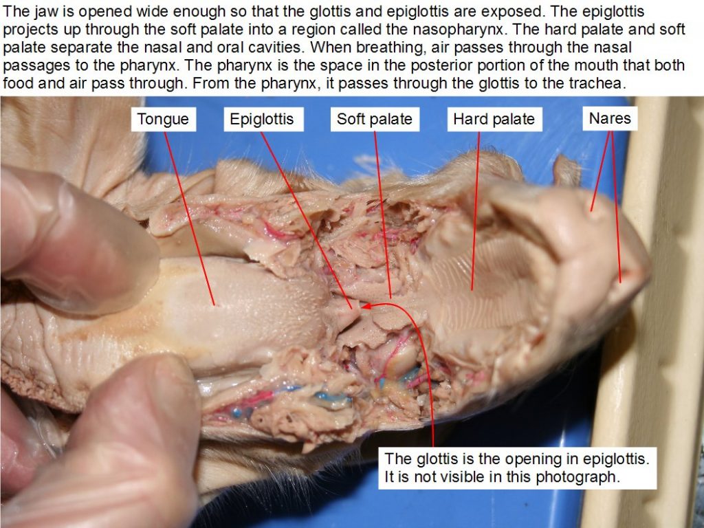
Carefully, peel the skin away from the incision in the neck region using a blunt probe (a needle or the point of scissors will do if a blunt probe is not available). Use the probe to peel away muscle tissue until the thymus gland on each side of the trachea is exposed.
Use a probe to separate the two lobes of the thymus gland and to further separate the musculature over the trachea. The thyroid gland is darker and lies between the posterior ends of the two lobes of the thymus gland.


Continue separating the tissue with a probe until the trachea and esophagus are exposed. The esophagus is dorsal to the trachea. The large hard structure attached to the trachea is the larynx. It contains the vocal cords.
In the photograph below, the heart and blood vessels of the neck region have been removed so that the trachea can be seen more clearly. You should not remove these structures yet because you will need to identify the blood vessels later in the dissection.
Below: esophagus, larynx, trachea, bronchus, lungs.
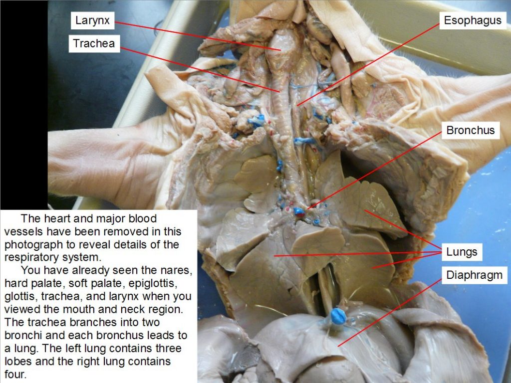
Respiratory System
Observe how the diaphragm attaches to the body wall and separates the abdominal cavity from the lung (pleural) and heart (pericardial) cavities (Photographs below). Contraction of the diaphragm forces air into the lungs.
You have already seen the pharynx, hard palate, soft palate, epiglottis, glottis, trachea, and larynx. Follow the trachea to where it branches into two bronchi and observe that each bronchus leads to a lung. The left lung contains three lobes and the right lung contains four. Each lung is located in a body cavity called a pleural cavity.


Digestive System
You have already seen how the esophagus leads from the pharynx through the neck region. Using a probe, trace follow the esophagus to the stomach. Identify the small intestine and large intestine. Find the posterior part of the large intestine called the rectum and observe that it leads to the anus. Locate the cecum, a blind pouch where the small intestine joins the large intestine.
Identify the liver. Lift the right lobe and find the gallbladder. This structure stores bile produced by the liver. Find the bile duct that leads to the small intestine. The pancreas is located dorsal and posterior to the stomach. It extends along the length of the stomach from the left side of the body (your right) to the point where the stomach joins the small intestine. Lift the stomach and identify this light-colored organ.
The spleen is an elongate, flattened, brownish organ that extends along the posterior part of the stomach ventral to (above) the pancreas.
The cecum is a blind pouch where the small intestine joins the large intestine. It houses bacteria used to digest plant materials such as cellulose. The cecum is large in herbivores but much of it has been lost during evolution in humans. The appendix in humans is the evolutionary remains of a larger cecum in human ancestors.


Circulatory System
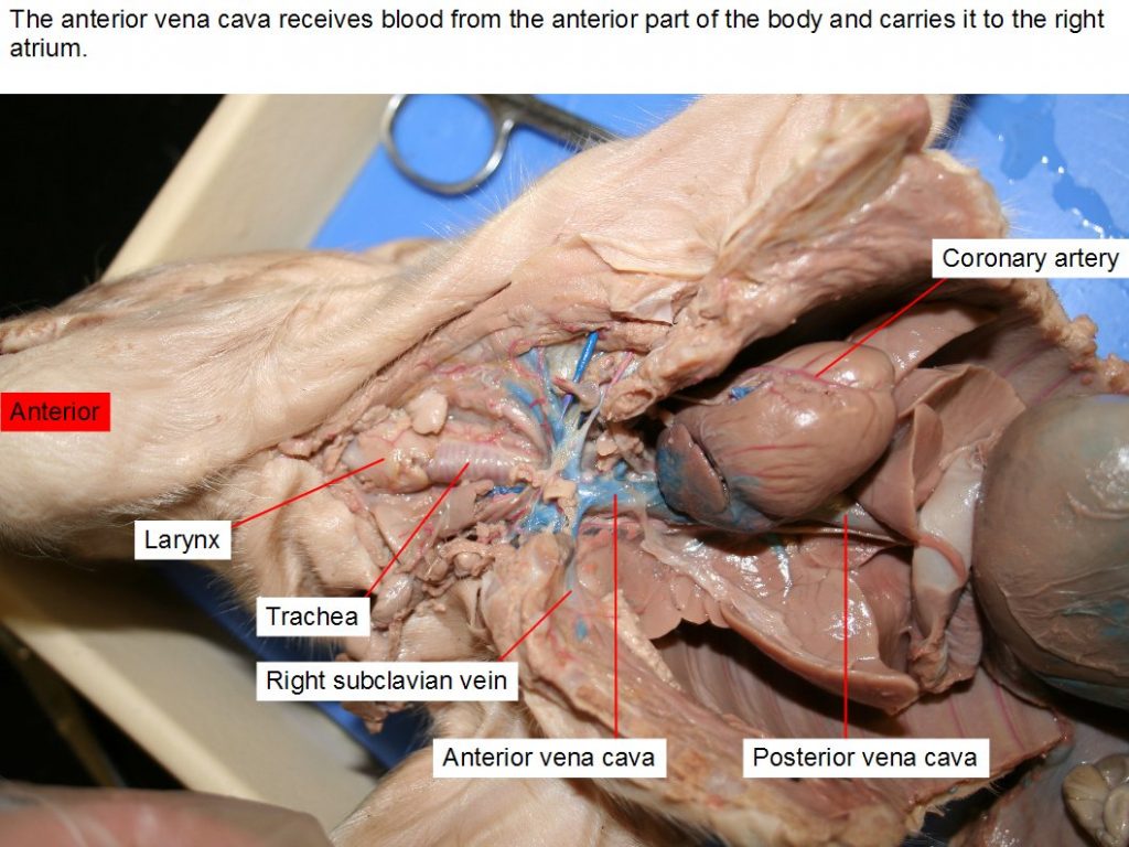
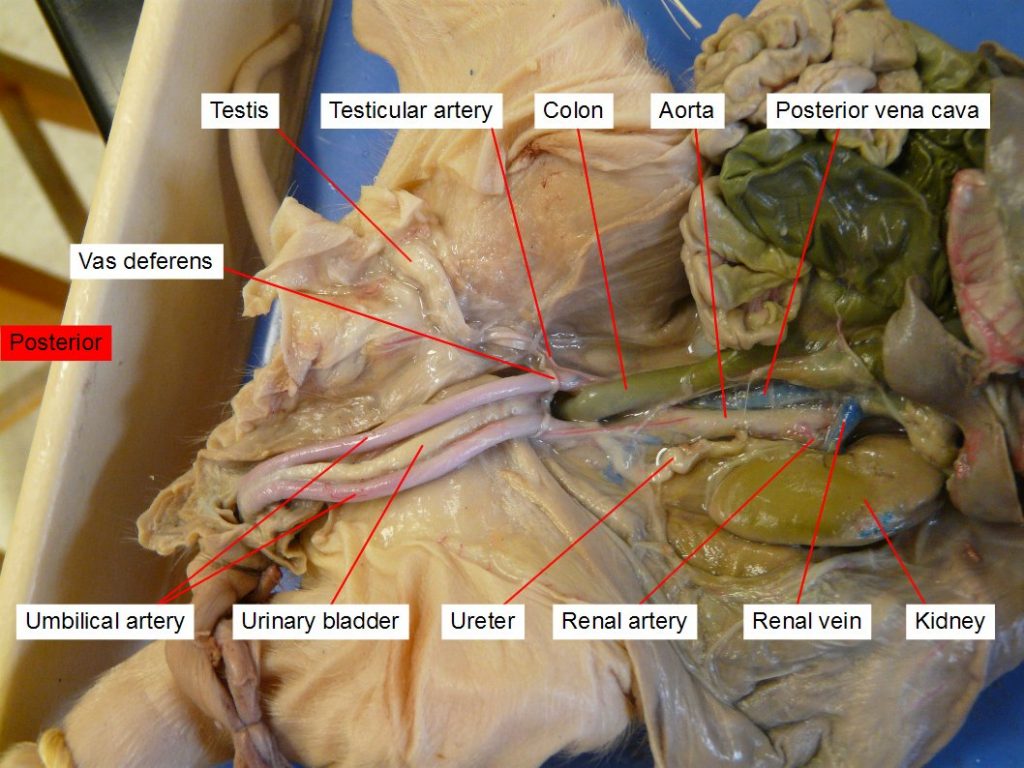
The diagrams below summarize the circulatory system of a mammal.

The drawing below shows some of the major arteries that carry blood to the body. Blood vessels that branch from the aorta carry blood to most of the body.
The pulmonary artery is capable of delivering a large amount of blood to the lungs but the lungs are not needed to oxygenate the blood of a fetus, so most of the blood is diverted to the aorta. This diagram shows that the ductus arteriosus connects the pulmonary artery to the aorta and diverts blood that would otherwise go to the lungs.
Shortly after birth, the ductus arteriosus closes and blood in the pulmonary artery goes to the lungs instead of the body.
Blood passes from the left ventricle through the aortic arch and aorta to the body. The first branch of the aorta is the brachiocephalic artery. The second branch is the left subclavian artery which goes to the left front leg. The right subclavian carries blood to the right front leg and the carotids carry blood to the head.


The pericardium is a membrane that surrounds the heart and lines the pericardial cavity. It contains a lubricating fluid and isolates the heart from body movements such as the expansion and contraction of the nearby pleural (lung) cavity.
To view details of the aortic arch, ductus arteriosus, and pulmonary artery, it will be helpful to remove the left lung. With the left lung removed, the heart can be pushed to the right side to reveal the aorta and other blood vessels shown in the diagram below.








Attributions for “Fetal Pig Dissection” MODIFIED from:
Original work by Michael J. Gregory, Clinton Community College / CC BY-NC-SA 3.0
