
Anatomy and Physiology
12 Nervous Tissue
 ʻIke i ke au nui me ke au iki.
Know the big currents and the little currents; being very well versed.
‘Ōlelo No‘eau, compiled by Mary Kawena Pukui, #1209
Introduction

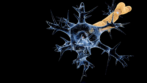
Figure 12.1 A drawing of pigeon Purkinje Cells, a type of neuron. Notice how the pattern looks like a branching waterway system.
How does the nervous system conduct the flow of information to the rest of the body? Communication within the nervous system is similar to that of ʻauwai (indigenous waterways). ‘Auwai serve to funnel water from a stream or spring in one area into farmlands or bodies of water in another. An entire ahupuaʻa could be supported by a single ʻauwai if it is properly built and maintained. If flow is interrupted farmers downstream from the blockage can’t water their plants. Similarly, neural impulses travel along unobstructed pathways connecting the to nearly every organ in the body. These neural impulses can spread quickly, flowing from a single nervous cell to potentially several subsequent cells. If these pathways are blocked, we lose control of the body parts downstream from the damaged nervous tissue. Just as water travels through ʻauwai networks, neural impulses move through complex pathways of the nervous system. ‘Auwai networks join together all the farmlands of an ahupua’a just as nervous sensory and motor communication connect the various organs of the body.
![]() Chapter Learning Outcomes
Chapter Learning Outcomes
- Differentiate between the CNS and PNS
- Distinguish between afferent, efferent, somatic and visceral neurons
- List nervous tissue cell types and their functions
- Detail the generation of graded and action potentials
- Illustrate the anatomy and physiology of a synapse
- Name the major neurotransmitters and chemical classification types
- Define the various nervous tissues and neural circuits
12.1 Central and Peripheral Nervous Systems (CNS & PNS)
![]() 12.1 Learning Outcomes
12.1 Learning Outcomes
- Relate the CNS and PNS to afferent and efferent signals
- Illustrate input, integration and output signals of the nervous system
- Compare and contrast the somatic and visceral nervous systems
The CNS and PNS

Figure 12.2 CNS and PNS are anatomically different parts of the nervous system. (OpenStax)
The nervous system is divided into the central nervous system (CNS) and the (PNS). The CNS consists of the brain and . The nervous tissue outside of the CNS is collectively called the PNS. The CNS and PNS coordinate in the following way:
- Detection of stimuli: Anything our body is able to detect is a and the begins in the PNS. Signals are generated and sent to the CNS. While we can be aware of some stimuli, such as sound, light, and odor, we are not aware of others, such as our blood pressure and pH.
- Integration of information: The CNS determines the response to stimuli which involves integrating multiple inputs and memories simultaneously. The response to any particular stimulus may be immediate, delayed or ignored completely.
- Response to stimuli: Emerging from the CNS, motor output signals target effectors (muscles and glands) in the PNS. These are motor output signals that stimulate tissues to control body movement and gland secretion.
- Homeostasis: The coordination between the CNS and PNS enables the body to maintain a healthy physiology. Notice how begins and motor function ends in the PNS, whereas the decision (integration) process occurs in the CNS. In some cases, organs are regulated by the CNS but have independent motor function. For example, cardiac muscle will contract without neural stimulation but the brain determines the contraction rate (aka the heart rate).


Figure 12.3 (a) Nervous System Detail (b) Stimuli detection starts in the PNS. Input signals are sent to the CNS via afferent (sensory) neurons. Integration occurs in the CNS which initiates an output signal via efferent (motor) neurons to target organs in the PNS.
Afferent (sensory) vs. Efferent (motor)
s are cells that send and receive signals in both the CNS and PNS. The neurons that send signals towards the CNS are afferent or s. They have sensory receptors that initiate signals sent to the brain or spinal cord. This is sensory input or sensation. The neurons in the CNS perform which is the process of assessing various sensory inputs and memories to determine an output response. The end result may be the contraction of muscles and/or secretion from glands. For this to happen, neurons must send signals away from the CNS to target organs in the PNS. These neurons are called efferent or motor neurons.

12.4 The relationship between somatic and visceral nervous systems.
Somatic vs. Visceral
The PNS consists of all nervous tissues outside the CNS. Remember, it consists of both sensory (input, afferent) and motor (output, efferent) neurons. Some of the s send sensory signals from the skin, joints, and muscles. These are s since they transmit sensory information about the body’s external environment, orientation and movement. s transmit sensory information from viscera or organs to the CNS. For example, if you feel nauseous, visceral sensory neurons in your stomach are sending signals to your CNS. There are also somatic and visceral s. s transmit signals to skeletal muscles. These signals are important in the movement and posture of the body. s, on the other hand, transmit signals to cardiac muscle, smooth muscle, and glands. These signals are important in the coordination of organ function to help maintain homeostasis. The nervous tissue controlling viscera is the since it generally works automatically (for example, you don’t have to remember to breathe or beat your heart). Opposite the ANS, the deals with conscious perception and both voluntary and reflexive movement of skeletal muscles.
Enteric Nervous System
There is a separate division of nervous tissue called the . The neurons in this division are embedded in the walls of gastrointestinal (GI) organs. These neurons function semi-autonomously to break down food into small absorbable molecules by coordinating smooth muscle contraction and secretion of digestive juices in the GI tract. As a result, we call the ENS the brain of the gut. Although the ENS functions somewhat on its own through intrinsic neural signals, it is also regulated by the extrinsic signals from the brain and spinal cord.
12.2 Cells of the Nervous System
![]() 12.2 Learning Outcomes
12.2 Learning Outcomes
- Detail the structure and function of neurons
- Differentiate the various neuroglia and neuron types
- Describe the process of myelination

Figure 12.5 Basic neuron structure. The dendrites and soma receive and then relay signals towards the s. There, the signal is transmitted to another neuron or tissue. (OpenStax).
Structure/Function of Neurons
Neurons
Neurons are the major functional cells of the nervous system. They detect stimuli, integrate information, conduct and relay signals, and initiate other tissue responses such as muscle contraction. Although there are different structural and functional types of neurons, they all share some common features. The , or just soma, is the cell body of the neuron which houses the . Extending from the cell membrane are cellular processes known as s and s. Dendrites and soma receive input from stimuli or other neurons and transmit electrical signals towards the axon. The axon then relays the electrical signal towards the which then branches into synaptic end terminals. The is the unique cytoplasm of axons that facilitate transmitting electrical signals. The ends of the terminals are often swollen puʻupuʻu (nodule) s or boutons. Here s are released which transmits the signal to other neurons or target organs such as skeletal muscles.
Between the cell body and the axon is a segment called the . This is where integration occurs within each neuron. If the signals received from the soma and dendrites are strong enough, the axon hillock sends a signal that conducts through the axon to the synaptic end terminals which triggers the release of neurotransmitters. Later in the chapter we will take a more detailed look at this process.
A is the neuron rough endoplasmic reticulum which is partially compartmentalized causing it to appear purple and granular when properly stained and viewed under a microscope. Nissl bodies continuously replace the neuron’s cell membrane as a normal process of growth and repair.
Some neurons can be quite long (over one meter!). Since large molecules and vesicles assemble in the neuron soma, an elaborate transport mechanism is needed to move them toward the axon terminal. is this active movement of substances, such as neurotransmitters, away from the cell body. A lot like how a train uses tracks, a motor protein called uses microtubules for anterograde transport. Some molecules in the terminals must be transported back to the soma for processing or recycling. Transport back to the soma, called , uses the same mechanism but employs a different motor protein called .
![]() Cultural Connection
Cultural Connection
Long before the Pali Highway was built, farmers on the windward side of Oʻahu transported their agricultural products to Honolulu by hiking a difficult trail over the Koʻolau mountains. They would sell their produce, buy any goods they needed and then return home. These farmers spent a lot of energy carrying heavy loads back and forth on this difficult journey. Similarly, kinesin and dynein use energy from ATP to move large substances to and from the axon terminals.

![]() Retrieval Practice
Retrieval Practice
After a thorough review of the figure and content about the structure of neurons, move away from your book and make a quick drawing. Label all the parts of the neuron. Add to those labels the functions of each of those parts. Check back in with your book and correct your drawing and labels. Take a little break to refresh your brain for optimal neuronal activity while you read the next section.
Neuroglia
s, or neuroglia, support neurons. There are 6 main types of glial cells each serving a distinct, vital role. Let’s read below the specific functions of each glial cell type.

Table 12.1 Structure/function/pathophysiology of Glial Cells (from Open Oregon State)
s (astro- = star; -cyte = cell) aid in the development of the . They have numerous processes (extensions) making them appear like a star. These extensions wrap around capillaries in the brain to regulate which blood compounds can enter the CNS. Another important function of astrocytes is their role as a potassium “buffer”. By absorbing or releasing potassium, astrocytes help to keep the concentration constant to maintain the electrical properties of neurons. Astrocytes also help remove excess neurotransmitters.

Figure 12.6 Neuroglial cells of the CNS. Astrocytes form the BBB, microglial cells provide immune function, oligodendrocytes form myelin and ependymal cells produce and regulate CSF. (OpenStax)
are small cells that wander around the CNS and replicate when there is inflammation caused by infection or damaged tissue. Similar to macrophages of the immune system, they remove debris and pathogens by engulfing and digesting substances in a process called phagocytosis.
s help to cushion and nourish the brain and spinal cord by producing a cerebrospinal fluid (CSF) that circulates throughout the CNS. Adjacent to capillaries, these cells are able to exchange waste products and nutrients to maintain CSF homeostasis.
s are large cells in the CNS with extensions that wrap around neuron axons forming a myelin sheath. Similar to the rubber coating on electrical wires, myelin helps insulate nervous signals. The glossy-white appearance of myelin is due to the high lipid content.

Figure 12.7 Neuroglial cells of the PNS. s wrap around neuron soma whereas wrap around the axon forming a myelin sheath. (OpenStax)
Satellite and Schwann cells are the two types of glial cells found in the PNS. Similar to oligodendrocytes, Schwann cells (aka neurolemmocytes) wrap around neuron axons in the PNS forming myelin sheaths. Satellite cells play similar nourishment and support roles as astrocytes but wrap around the neuron cell body.
![]() Clinical Application
Clinical Application
Multiple sclerosis is a condition where there is progressive demyelination of neurons in the CNS due to the loss of oligodendrocytes. The loss of myelin disturbs the conduction of nervous signals causing impairment of sensory and motor function. Multiple sclerosis is a progressive disorder meaning it gets worse over time. The loss of myelin on neurons in the PNS is known as . As with multiple sclerosis, this disorder is characterized by impaired sensation and muscle weakness. However, unlike multiple sclerosis, this disorder often resolves spontaneously.


Guillain-Barré Syndrome: See motion illustration: https://commons.wikimedia.org/wiki/File:Guillain-barr%C3%A9_syndrome_-_Nerve_Damage.gif

Figure 12.8 Oligodendrocytes and Schwann form myelin sheaths. (a) Notice the nucleus (upper right corner) of the myelin forming cell stays on the outside of the sheath as it wraps around the axon. (b) EM micrograph provided by the Regents of University of Michigan Medical School. 1,460,000x magnification (OpenStax)
Both oligodendrocytes (CNS) and Schwann cells (PNS) produce sheaths by wrapping around axons to form layers of myelin in a process known as myelination. A single oligodendrocyte can myelinate axons of multiple neurons simultaneously whereas a single Schwann cell often just myelinates a small part of one axon. The insulating effects of myelination allow neural signals to be transmitted more quickly.
The myelin sheath is not continuous along the axon. There are gaps between sheaths where the neuron plasma membrane is exposed. These regions are called (aka neurofibril nodes). In addition, some axons are heavily myelinated whereas others are either lightly or unmyelinated. Even in unmyelinated neurons, oligodendrocytes and Schwann cells may cover parts of the axon but with too few layers for an insulating effect.

Figure 12.9 Types of Neurons. Multipolar neurons have many dendrites extending from the soma and one long axon with many terminal branches (a). have one dendrite and axon each but with potentially several branches (b). have one axon with branches of dendrites and terminals at opposite ends (c). (Open Oregon State)
Neuron Types
There are three main types of neurons classified by the number of cellular processes that extend from the soma. The most common type are multipolar neurons. These neurons have several dendrites and a single axon extending from the cell body. Examples include motor and interneurons which constitute most of the brain and spinal cord.
The second type are bipolar neurons. These neurons have just two processes attached to the soma; a single dendrite and a single axon. Despite having just one of each, both the axon and dendrite may have many branches. Bipolar neurons function in some of the special sense organs including the retina of the eye and the olfactory epithelium in the nose.
Finally, unipolar neurons have a single process extending from the cell body. This process connects directly to the axon. Instead of being attached to the cell body, dendrites of unipolar neurons are attached to the end of the axon opposite the axon terminal. Unipolar neurons are involved in sensation pathways.
12.3 Membrane Potentials
![]() 12.3 Learning Outcomes
12.3 Learning Outcomes
- Distinguish between the various pumps and ion channels
- Employ Ohm’s law to explain polarization/ depolarization of the neuron membrane
- Relate the resting membrane potential to depolarization and hyperpolarization
- Differentiate graded and action potentials
- Compare and contrast continuous versus saltatory conduction and the fiber types
Overview
Neurons relay electrical signals from their dendrites and soma toward the axon terminal. Our ability to appreciate music, learn new concepts, rest in a hammock, and body surf at Manini’owali Beach all depend on these impulses! To generate and relay electrical signals, membrane proteins control the movement of ions across the neuron membrane. Consistent with the concept of form dictating function, the kūkulu ʻana (arrangement, organization) of these membrane proteins that determines how signals are received and transmitted.

Figure 12.10 Sodium-potassium (Na+/K+) pumps move 3 Na+ ions out of the cell for every 2 K+ ions brought in.
Pumps and Channels
As you may recall from previous chapters, membrane protein pumps concentrate ions by moving them in or out of the cell. Concentrating ions requires cellular energy via ATP. Sodium-potassium (Na+/K+) and are the most common ion pumps. Continuously and throughout the neuron, Na+/K+ pumps move three Na+ ions out for every two K+ ions brought in whereas Ca++ pumps simply move Ca++ ions out. Because pumps move ions in the direction of greatest concentration, it is often described as moving ions up a concentration gradient.
 |
 |
 |
 |
Figure 12.11 Leakage, ligand-gated, voltage-gated, and mechanically-gated ion channels. Leakage channels randomly open and close, essentially allowing a slow but continuous diffusion of ions (a). Ligand-gated ion channels open only upon neurotransmitter binding (b). Voltage-gated ion channels open when the of the membrane changes (c). Mechanically-gated channels open when a physical force is applied (d). Channel subtypes exist for each specific ion.
Channels are proteins with pores that allow ions to diffuse down their concentration gradient. Diffusion does not require energy input. There are four types of channels: leakage, ligand-gated, voltage-gated, and mechanically-gated channels. s (a.k.a. passive channels) are always open allowing for continuous diffusion. (a.k.a. chemical channels) are usually closed. They open when a chemical messenger, such as a neurotransmitter binds to them. s are typically closed as well. They open when the electrical conditions of the membrane change. Mechanically-gated channels open when some physical force is applied such as those on neurons involved in the sensation of touch.
Remember, a major theme in anatomy and physiology is that structure is related to function. This concept applies to the distribution of channels across the neuron membrane. In soma and dendrite membranes, we find ligand or s for sodium, potassium and chloride. These open in response to stimuli or neurotransmitters as a way of receiving a signal. In the axon region, there are many voltage-gated channels for sodium and potassium. These channels propagate that signal down the axon. In the axon terminals there are voltage-gated calcium channels. Opening these stimulates neurotransmitter release which relays the signal to the following neuron(s).
![]() Local Issue
Local Issue
On the island of Oʻahu, the Navy built the Red Hill Underground Fuel Storage Facility in 1943. In 2021, this facility leaked several thousand gallons contaminating a major drinking water aquifer. It took several months before it was detected and action taken to prevent nearby residents consuming the contaminated water. Thousands of people were affected with neurological issues being a major complaint. Headaches, nausea and seizures are a few of the common neurological problems caused by exposure to gasoline regardless if it is inhaled, ingested or injected. This volatile compound escapes the body via evaporation through the lungs. This causes pneumonitis and subsequent hypoxia. Lack of oxygen disrupts normal neuron physiology resulting in these symptoms.

Red Hill as viewed from Hālawa Valley

Figure 12.12 Membrane Potential. The difference in electrical charge between the inside and outside of the cell membrane is a potential energy and measures around -70mV when the cell is at rest (ion gates are closed).
Resting Membrane Potential
Now let’s take a closer look at the details of how neurons receive, conduct and relay signals. The movement of charged particles is electrical energy. The most familiar example is the movement of electrons across metal wires in electronic devices. However, the movement Na+ or K+ is also a form of electricity. When we think about electricity there are three important characteristics to keep in mind: voltage, current, and . Voltage (aka potential energy) is the difference in electrical charge between two areas. In the case of the human cell, there is an electrical charge difference between the outside and inside of the membrane. This is due to Na+/K+ pumps continuously moving three positive charges (Na+ ions) out of the cell for every two positive charges (K+ ions) brought in. Remember there are Na+ and K+ leakage channels too but these only allow a slow diffusion of ions. In addition, cells have a high concentration of negatively charged intracellular proteins. The difference in electrical charge is voltage and across the neuron plasma membrane this measures around -70mV. This is referred to as the . When gated ion channels open ions quickly diffuse creating a current which changes the from its resting state. Resistance is due to the physical properties of the membrane and channels that limits the diffusion rate of ions.
Current and Ohm’s Law
Whereas voltage is the separation of charged particles, current is the movement of charged particles. Here in Hawaiʻi, most of us are familiar with au kai (ocean current) that describes the movement of water. The opposition to movement is resistance. For instance, if a Na+ gated channel were to open large amounts of Na+ ions would quickly diffuse into the cell. That movement generates a current. However, the gate size limits how quickly Na+ can diffuse in and therefore imparts a resistance. The relationship between current, voltage and resistance is called .
Ohm’s Law:
I=V/R
where I = current, V = voltage and R = resistance.

Figure 12.13 Polarization, depolarization and hyperpolarization of a localized membrane depends on the specific gated ion channels that open. Image c voltmeter should read -90mV or something less than -70.
Membrane Polarity
When a cell has established a RMP the membrane is described as because one side is negatively charged relative to the other. When Na+ gates open, sodium rushes into the cell and the membrane becomes less polar or . It will be helpful to associate the RMP with polarization of the membrane and the opening of Na+ gated channels with depolarization of the membrane. When Na+ gates close, the Na+/K+ pumps will eventually the membrane to the RMP state. When K+ or Cl- gates open the membrane s, it becomes less than the -70mV RMP. Remember, depolarization and hyperpolarization begins where ion gates open and only spreads beyond the localized membrane if other gates are triggered to open as well.

Figure 12.14 Graded Potentials Some stimuli may only depolarize the membrane a small amount due few ligand-gated Na+ channels being opened and only for a short time. These are stimuli. Other stimuli may hyperpolarize the membrane due to opening K+ or Cl- gated channels. Only stimuli that depolarize the membrane potential above -55mV may initiate the opening of voltage-gated Na+ channels generating an .
Graded (local) Membrane Potential
At rest, neurons are polarized around -70mV. When dendrite and soma ligand-gated Na+ channels are stimulated to open, the membrane begins to depolarize but just near the opened gates. The membrane may depolarize a little bit or a great amount depending on the number of gates opened and the frequency of stimulation. Because these disruptions may be weak or strong, depolarization signals in dendrites and soma are called s. Because graded potentials are isolated around the opened gates, they are also referred to as s. Graded potentials initiated by sensory receptors are called receptor or s. When graded potentials are strong enough to depolarize the membrane beyond a certain level, it may open voltage-gated Na+ channels in the axon hillock. If this occurs, we have created an action potential!
Action Potential
How are action potentials generated from graded potentials? Opening of dendrites and soma Na+ gated channels cause the membrane voltage to rise from the RMP of -70mV. When the graded potentials reach a level called the (around -55mV), nearby voltage-gated Na+ channels may open. This causes more Na+ to rush in depolarizing the membrane further away from where the graded potential was initially generated. If enough Na+ rushes in, the graded potential may open voltage-gated Na+ channels in the axon hillock. Here, in the axon hillock and for the rest of the axon, voltage-gated Na+ channels are close enough to one another so that if one gate opens other nearby gates also open. This of the depolarization signal down the length of the axon towards the terminus is an action potential. Whereas graded potentials may or may not open adjacent channels, as long as one voltage-gated Na+ channel opens in the axon hillock all will open in sequence across the axon. This is why action potentials are described as “all or nothing” responses. This process begins in the axon hillock which is often referred to as the “trigger point.” If graded potentials don’t trigger an action potential it is called a subthreshold stimulation.
![]() Deep Dive
Deep Dive
Local anesthetics, such as lidocaine, that dentists apply when filling a cavity in a tooth, block voltage-gated sodium channels and therefore block action potentials that carry pain information. Can you visualize how that works? Think about that next time you visit your dentist!


Figure 12.15 The RMP of -70mV (1) rises when Na+ gates open (2). If threshold is reached, the membrane potential may reach as high as +30mV (3). Then, voltage-gated K+ channels open which repolarizes the membrane (4). These gates stay open long enough to cause a temporary hyperpolarization (5) before returning to the RMP (6).
When voltage-gated Na+ channels open the membrane potential reaches around +30mV which then triggers the gate to close. During this time, the much slower voltage-gated K+ channels open. As a consequence, potassium quickly diffuses out of the neuron. With positive charges now leaving the neuron, the membrane potential starts becoming less positive in a process called repolarization. Voltage-gated K+ channels also stay open for a little longer. As a result, potassium continues to diffuse out of the neuron causing a brief hyperpolarization. Once these close, the RMP is reestablished by the Na+/K+ pump returning the sodium and potassium to outside and inside of the membrane, respectively. The field of science researching the behavior of membranes and membrane channels is called electrophysiology.

Figure 12.16 Absolute and Relative refractory periods. During absolute refractory, the neuron cannot be stimulated to depolarize again. During the relative a stronger stimulus is needed to induce depolarization.
Because it takes time for K+ gates and Na+/K+ pumps to repolarize the membrane, a neuron cannot be stimulated again for about 1 ms. This period is known as the . This period ensures the action potential is directed towards the axon terminals. There is also the where an action potential can be initiated as long as the stimulus is very strong and much greater than the normal stimulus required to generate an action potential. The relative refractory period occurs right after the absolute refractory period when the voltage-gated K+ channels are still open but the voltage-gated Na+ channels have reset and are ready to open again.
![]() Food and Environment
Food and Environment
Have you ever heard about the pufferfish? These fish have the ability to inflate themselves when they are threatened. But this is probably not the most interesting feature of this fish. Pufferfish have a toxic substance called tetrodotoxin which is produced by the symbiotic or infecting bacteria living in its liver. This substance is a neurotoxin that blocks sodium channels in the neurons, interrupting the proper functioning of action potentials. So, think about that when choosing the fish for your next sushi meal!
 |
 |
 |
Figure 12.17 Continuous and Saltatory Conduction Gated ion channels are required throughout the axon to propagate continuous action potentials. Where the axon is myelinated, the electric signal jumps from one node of Ranvier to another. is much faster than continuous.
Propagation
An action potential is generated at the axon hillock and is conducted along the axon towards the terminus in a process called propagation. In unmyelinated axons, this is a result of numerous sodium and potassium voltage-gated channels close enough to one another so that if one opens nearby gates will also open. Because the channels open in a sequential manner, this type of action potential is described as a continuous conduction and is characteristic of unmyelinated axons. On the other hand, in myelinated axons, the voltage-gated channels for sodium and potassium open are only present where myelin is absent; the areas known as the nodes of Ranvier. In these axons, action potential propagation is known as saltatory conduction. Saltar means to lele (jump, skip). The transmission of the action potential in myelinated axons is much faster than in unmyelinated axons because it doesn’t rely on the opening and closing of ion gates.

Figure 12.18 Fibers Type A fibers have the largest diameters and are heavily myelinated. Type B fibers are intermediate in size and myelination. Type C fibers are the thinnest and have no myelin sheath.
are bundles of axons classified according to their diameter and degree of myelination. There are three types of fibers: A, B, and C. Motor neurons that innervate skeletal muscles have . These fibers have large diameters, are highly myelinated, and therefore conduct action potentials in a very fast way. are lightly myelinated with intermediate diameters. Finally, are unmyelinated and have the smallest diameters. Conduction of action potentials in type C fibers is the slowest.
12.4 Synapses and Neurotransmitters
![]() 12.4 Learning Outcomes
12.4 Learning Outcomes
- Outline the events of signal transmission at chemical synapses
- List the structures and functions of various neurotransmitters
- Compare and contrast ionotropic and metabotropic receptors
- Describe neurotransmitter degradation and reuptake

Figure 12.19: The Synapse Synapses include the axon terminal region, the adjacent membrane of the and the cleft in between.
Synapse Structure and Function
The area where two neurons come together is a . It consists of the synaptic end terminal region of the known as the presynaptic membrane, a narrow space called the , and the adjacent postsynaptic membrane of the postsynaptic neuron.

Figure 12.20 Chemical and Electrical Synapses Chemical synapses involve the release of neurotransmitters. Electrical synapses have ion channels to stimulate the postsynaptic cell directly.
When an action potential reaches the synaptic end terminal, it causes voltage-gated Ca++ channels to open. This triggers a cascade of events leading to the release of neurotransmitters into the synaptic cleft. The neurotransmitters then diffuse across the cleft, binding receptors in the postsynaptic membrane causing graded potentials in muscles, glands or other neurons. Since a neurotransmitter, a chemical, causes these gates to open they are chemical synapses. In some cases the action potential of one neuron directly depolarizes another without using a neurotransmitter. These are s. Either way, the effect on postsynaptic neurons is referred to as the .
 |
 |
Figure 12.21 Neurons can synapse in a variety of ways aside from axosomatic.
Though we typically think of synapses as being constructed of the axon terminal of a presynaptic neuron and the dendrites of a postsynaptic neuron, sometimes neurons synapse in other ways. There are axo-axonic, dendro-dendritic, somato-dendritic, dendro-somatic, somato-somatic among other types of synapses.
| System | Cholinergic | Amino acids | Biogenic amines | Neuropeptides |
| Neurotransmitters | Acetylcholine | Glutamate, glycine, GABA, aspartate | Serotonin (5-HT), dopamine, norepinephrine, (epinephrine) | Met-enkephalin, beta-endorphin, VIP, Substance P, etc. |
| Receptors | Nicotinic and muscarinic receptors | Glu receptors, asp receptors, gly receptors, GABA receptors | 5-HT receptors, D1, and D2 receptors, α-adrenergic and β-adrenergic receptors | Receptors are too numerous to list, but are specific to the peptides. |
| Elimination | Degradation by acetylcholinesterase | Reuptake by neurons or glia | Reuptake by neurons | Degradation by enzymes called peptidases |
| Postsynaptic effect | Nicotinic receptors cause depolarization. Muscarinic receptors can cause both depolarization or hyperpolarization depending on the subtype. | Glu and Asp receptors cause depolarization. Gly and GABA receptors cause hyperpolarization. | Depolarization or hyperpolarization depends on the specific receptor. For example, D1 receptors cause depolarization and D2 receptors cause hyperpolarization. | Depolarization or hyperpolarization depends on the specific receptor. |
Table 12.2 Neurotransmitter chemical classification
Neurotransmitter Classes and Receptors
There are different chemical classes of neurotransmitters. Some classes include multiple neurotransmitters with unique effects. In other cases, a single neurotransmitter may have varied effects, depending on what synapses it is released.
is the most common neurotransmitter in the body. The adjective cholinergic describes this neurotransmitter and the receptors it binds. Acetylcholine can excite or inhibit depending on which receptors the target cell has. There are two general types of cholinergic receptors; s and muscarinic receptors. Both of these bind ACh but nicotinic receptors can also bind nicotine and muscarinic receptors muscarine. Nicotinic receptors are excitatory whereas muscarinic receptors can be excitatory or inhibitory depending on the subtype. ACh is involved in all NMJs, most ANS pathways among other synapses.
Amino acid neurotransmitters include , , , and . Glutamate and aspartate are the most common excitatory neurotransmitters in the brain and spinal cord, respectively. On the other hand, GABA and glycine are the most common inhibitory neurotransmitters in the brain and spinal cord. Aside from their predominant roles, each of these also have other effects dictated by the specific receptors of the various synapses.
neurotransmitters include amine groups but do not have carboxylic acids. This class of neurotransmitters includes serotonin (aka 5-hydroxytryptamine or 5-HT), dopamine, norepinephrine, and epinephrine. As with the amino acid neurotransmitters, the effects of these vary depending on the receptor bound.
Finally, neurotransmitters are a chain of 2-40 amino acids. This class of molecules include enkephalin, substance P, cholecystokinin, beta-endorphin, and many others. These neurotransmitters tend to be more localized with more specialized functions.

Figure 12.22 Receptor Types Ionotropic receptors are ion gates that open upon neurotransmitter binding (a). Metabotropic receptors activate G proteins leading to the productions of 2nd messengers (b). These molecules can have a multitude of effects including the opening of ion channels.
Receptor types, up/down regulation and pharmacology
Generally speaking, neurotransmitter receptors can be categorized as ionotropic or metabotropic. Ionotropic receptors are ligand-gated channels. When the neurotransmitter binds, a gate opens allowing one or more types of ions through. Ionotropic receptors function quickly because of the direct mechanism of action. The net effect depends on the specificity of the ion channel. For example, a class of glutamate ionotropic receptors called AMPA is selectively permeable to Na+. Therefore, when glutamate binds to this receptor type it leads to a rapid depolarization. Another binds GABA and is selectively permeable to Cl-. When GABA binds to this receptor, it leads to a rapid hyperpolarization.
s have an indirect mechanism of action and are therefore much slower. Here, the second messenger system is employed. This means that before an ion gate is induced to open a second messenger must first be activated. Most commonly, the neurotransmitters bind which increase or decrease a second messenger such as cyclic adenosine monophosphate (cAMP) or cyclic guanosine monophosphate (cGMP). Some additional effects of second messenger signal transduction pathways include regulation of metabolism and gene expression. Therefore the effects of neurotransmitters binding metabotropic receptors, although slower, can have more diverse and long-lasting effects. Examples of metabotropic receptors include the muscarinic ACh receptors and the adrenergic receptors for norepinephrine.

Figure 12.23 Synapse During Reuptake Neurotransmitter effect regulated by enzymatic degradation within the synapse or by for recycling or intracellular degradation.
Neurotransmitters can quickly excite or inhibit a target cell but then, for optimal control, the signal needs to be quickly stopped. There are two main mechanisms to prevent neurotransmitters from continuing to activate receptors. The first is . For example the enzyme breaks down ACh into acetic acid and choline. Choline is then transported back into the cell for use in the synthesis of more ACh. Another mechanism of inactivation is reuptake which is the transport of neurotransmitters back into the presynaptic neuron where it is internally degraded. An example of this mechanism is the reuptake of serotonin which is transported back into the presynaptic neuron where it is degraded by the enzyme monoamine oxidase (MAO). A practical application of this is the treatment of depression. Since levels of serotonin are lower in patients with depression they can be treated with , a medication that inhibits the reuptake pumps that transport serotonin back into the neuron. Another type of medication that can be used is a that inhibits the enzyme that breaks down serotonin. Either of these medications would increase serotonin levels and subsequent effects.
12.5 Neuronal Integration and Neural Circuits
![]() 12.5 Learning Outcomes
12.5 Learning Outcomes
- Explain neuronal integration
- Relate ganglia, nerves, nuclei, tracts and gray and white matter
- Illustrate the various types of neural circuits

Figure 12.24 Postsynaptic Potential Summation The axon hillock is where summation of all EPSPs and IPSPs occur. Graded excitatory and inhibitory potentials are continuously disrupting the RMP (A). If the threshold of -55 mV is reached an action potential occurs (B).
Integration
When neurotransmitters bind a receptor, ion gates open causing the cell to depolarize or hyperpolarize. Depolarization signals may generate action potentials, therefore these are called s (EPSPs). Hyperpolarizing signals are called s (IPSPs). Graded potentials weaken as they travel away from the receptor. In order to trigger an action potential within the axon hillock, many EPSPs must be received simultaneously or one receptor must be stimulated repeatedly. The axon hillock adds (s) together all EPSPs and IPSPs. If the summation of all signals is above threshold then an action potential will be generated.
occurs when adjacent synapses are activated simultaneously. occurs when a single synapse is rapidly activated over and over.
Some synapses are inhibitory, some are excitatory, but the net effect is due to the summation of all of them at the axon hillock. There may be thousands of synapses covering the dendrites and soma of a single neuron. If summation causes the membrane potential to reach the threshold, an action potential is triggered. However, if summation of all graded potentials only reaches a subthreshold level an action potential is not triggered. This is determined by the rate of firing of neurons at each synapse, the number of synapses involved but also the proximity of each synapse to the hillock. Graded membrane potentials generated closer to the axon hillock degrade less than those further away. Therefore, these synapses generate signals that are more heavily weighted during summation. Despite this complexity, spatial and temporal summation ensures that neurons ultimately act like a switch; either an action potential is created or it isn’t.

Figure 12.25 Tissue Terminology of the CNS and PNS. In the CNS, a group of neuron soma are referred to as gray matter or a nucleus, whereas in the PNS it occurs as a (swelling). Axon regions in the CNS are or s. In the PNS, axons bundle into nerves.
Neuron Organization
Both in the CNS and PNS, neurons tend to organize such that soma and axons usually group separately. In the CNS, groups of neuron soma are or nuclei. Typically, gray matter refers to superficial regions and nuclei deeper foci. Often neuron soma grouped in a nucleus work together for a common purpose. These s control various functions such as breathing, heart rate, and most other bodily functions. Grouped axons in the CNS are white matter or tracts. In the PNS, grouped soma are ganglia whereas grouped axons are called nerves.

Figure 12.26 Circuit Types. Converging, diverging, reverberating and parallel-after-discharge circuits.
Circuit Types
Multiple nuclei coordinate together to achieve some complex bodily functions. The sequence of neuron interaction is referred to as a circuit. Although some are complex, we can categorize circuits as either converging, , reverberating or parallel-after-discharge circuits. Converging circuits involve many sources of input which then act upon a single output neuron. For example, the rate and depth of breathing is influenced by many sensory inputs such as blood pH, emotions, and pain among others.
Diverging circuits are the opposite. Here, a single nucleus could send signals to many output targets. For example, the “fight or flight” sympathetic nervous system nuclei can send signals to increase heart and breathing rate and dilate the airways, pupils of the eye, and blood vessels. The divergence of signals from nuclei such as this can involve thousands of different targets.
s are for rhythmic functions that will continue until there is an inhibitory signal. For example, they are involved in maintaining breathing patterns even while sleeping.
Parallel-after-discharge circuits are a combination of converging and diverging circuits. First, a signal can diverge from a nucleus with multiple pathways but then converge onto another nucleus. The diverging pathways may be of different lengths and numbers for neurons and therefore converge at different times. These are thought to play a role in more complex functions such as higher order thinking.

Chapter Summary

Key Terms
absolute refractory period
period time during an action period when another action potential cannot be generated because the voltage-gated Na+ channel is inactivated
acetylcholine (ACh)
one of the most common and well studied neurotransmitters, it plays roles in all NMJs and many synapses of ANS pathways
acetylcholinesterase (AChE)
an enzyme that breaks down ACh within synapses to regulate its effect
action potential
change in voltage of a cell membrane in response to a stimulus that results in
transmission of an electrical signal; unique to neurons and muscle fibers
afferent neuron
neuron that sends nervous signals towards the brain and/ or spinal cord
anterograde transport
the transport of substances from neuron soma toward axon terminals
aspartate
a common excitatory amino acid neurotransmitter of the spinal cord among other roles
astrocyte
glial cell type of the CNS that provides support for neurons and maintains the blood-brain barrier
autonomic nervous system (ANS)
functional division of the nervous system that is responsible for homeostatic reflexes that coordinate control of cardiac and smooth muscle, as well as glandular tissue
axon
single process of the neuron that carries an electrical signal (action potential) away from the cell body toward a target cell
axon hillock
tapering of the neuron cell body that gives rise to the axon
axon terminal
end of the axon, where there are usually several branches extending toward the target cell
axoplasm
cytoplasm of an axon, which is different in composition than the cytoplasm of the neuronal cell body
biogenic amine
class of neurotransmitters that are enzymatically derived from amino acids but no longer contain a carboxyl group such as serotonin, dopamine, norepinephrine, and epinephrine
bipolar neurons
shape of a neuron with two processes extending from the neuron cell body—the axon and one dendrite
blood-brain barrier (BBB)
physiological barrier between the circulatory system and the central nervous system that establishes a privileged blood supply, restricting the flow of substances into the CNS
brain
the large organ of the CNS composed of white and gray matter, contained within the cranium and continuous with the spinal cord
calcium (Ca++) pumps
proteins that pump Ca++ ions out of the cytoplasm to the ER or extracellular space
central nervous system (CNS)
anatomical division of the nervous system located within the cranial and vertebral cavities, namely the brain and spinal cord
cerebrospinal fluid (CSF)
circulatory fluid within the CNS that is produced by ependymal cells in choroid plexuses
chemical synapse
connection between two neurons, or between a neuron and its target, where a neurotransmitter diffuses across a very short distance
cholinergic
system neurotransmitter system of acetylcholine, which includes its receptors and the
enzyme acetylcholinesterase
continuous conduction
slow propagation of an action potential along an unmyelinated axon owing to voltage-gated Na+ channels located along the entire length of the cell membrane
converging circuit
a neuron circuit by which several input neurons converge onto one neuron
current
the movement of charged particles from one area to another
dendrite
one of many branchlike processes that extends from the neuron cell body and functions as a contact for incoming signals (synapses) from other neurons or sensory cells
depolarized
change in a cell membrane potential from rest toward zero effector protein enzyme that catalyzes the generation of a new molecule, which acts as the intracellular mediator of the signal that binds to the receptor
diverging
a neuron circuit whereby one input neuron acts on several other neurons
dynein
a motor protein involved in neuronal retrograde transport
efferent neuron
neuron that sends nervous signals away from the brain and/ or spinal cord
electrical synapse
connection between two neurons, or any two electrically active cells, where ions flow
directly through channels spanning their adjacent cell membranes
enteric nervous system (ENS)
neural tissue associated with the digestive system that is responsible for nervous control through autonomic connections
enzymatic degradation
a process of regulating neurotransmitter effect by breaking it down soon after release into the synaptic cleft
ependymal cell
glial cell type in the CNS responsible for producing cerebrospinal fluid
excitatory postsynaptic potential (EPSP)
graded potential in the postsynaptic membrane that is the result of depolarization and makes an action potential more likely to occur
gamma-amino-butyric-acid (GABA)
a common inhibitory amino acid neurotransmitter of the brain among other roles
ganglion (pl. ganglia)
localized collection of neuron cell bodies in the peripheral nervous system
gated property of a channel that determines how it opens under specific conditions, such as voltage change or physical deformation
generator potential
graded potential in dendrites of a unipolar neuron which generates an action
potential in the initial segment of that cell’s axon
glial cell (aka neuroglia)
one of the various types of neural cells responsible for maintenance of nervous tissue including the support of neurons
glutamate
a common excitatory amino acid neurotransmitter of the brain among other roles
glycine
a common inhibitory amino acid neurotransmitter of the spinal cord among other roles
G-protein coupled receptors
receptors that are associated with cytoplasmic G-protein, a GTP hydrolase, that physically moves from the receptor to a effector protein resulting in the production of cAMP or cGMP (aka second messengers)
graded potential
change in the membrane potential that varies in size, depending on the size of the
stimulus that elicits it
gray matter
regions of the nervous system containing cell bodies of neurons with few or no myelinated axons; actually may be more pink or tan in color, but called gray in contrast to white matter
Guillain-Barré Syndrome
a pathologic condition caused by demyelination of neurons in the PNS
hyperpolarize
state of the cell membrane when it polarizes more than the RMP
inhibitory postsynaptic potential (IPSP)
hyperpolarizing graded potentials in the postsynaptic membrane
integration
nervous system function that combines sensory perceptions and higher cognitive functions (memories, learning, emotion, etc.) to produce a response
ionotropic receptor
an ion channel gate that opens by neurotransmitter binding
kinesin
a motor protein involved in neuronal anterograde transport
leakage channel
ion channel that opens randomly and is not gated to a specific event, also known as a non-gated channel
ligand-gated channels
ion channel that opens upon neurotransmitter binding
local potential
change in membrane potential that is limited to the area nearby an open ion gate
mechanically gated channel
ion channel that opens when a physical event directly affects the structure of the protein
membrane potential
distribution of charge across the cell membrane, based on the charges of ions
metabotropic receptor
neurotransmitter receptor that involves a complex of proteins that cause
metabolic changes in a cell
microglia
cell type in the CNS that serves as the resident component of the immune system
monoamine oxidase inhibitor (MAOI)
a drug prescribed to serotonin deficient patients to potentiate the neurotransmitter effects by reducing intracellular enzymatic degradation
motor neuron
neuron that send action potentials towards muscles and glands for body movement and regulating physiology
multipolar neurons
shape of a neuron that has multiple processes—the axon and two or more dendrites
multiple sclerosis
a pathologic condition caused by progressive demyelination of neurons in the CNS
muscarinic receptor
type of acetylcholine receptor protein that is characterized by also binding to
muscarine and is a metabotropic receptor
myelin
lipid-rich insulating substance surrounding axons of some neurons
myelination
process of producing layers of myelin around neuron axons
myelin sheath
multiple layers of insulating myelin of some neuron axon regions allowing for faster transmission of electrical signals
nerve
cord-like bundle of axons located in the peripheral nervous system that transmits sensory input and response output to and from the central nervous system
nerve fiber
bundles of axons classified as either type A, B, or C fibers according to their diameter and degree of myelination
neuron
neural tissue cell that is primarily responsible for generating and propagating electrical signals into, within, and out of the nervous system
neuronal pool
a group of neurons within a nucleus serving a common function
neuropeptide
neurotransmitter of short chain (2-40) amino acids
neurosoma (aka soma)
in neurons, that portion of the cell that contains the nucleus; the cell body, as opposed to the cell processes (axons and dendrites)
neurotransmitter
chemical signal that is released from the synaptic end bulb of a neuron to cause a
change in the target cell
nicotinic receptor
type of acetylcholine receptor protein that is characterized by also binding to
nicotine and is an ionotropic receptor
Nissl body
rough ER of neurons which appears purple and granular when viewed under a microscope
nodes of Ranvier (aka neurofibril nodes)
gaps between two myelinated regions of an axon
nucleus (pl. nuclei)
in the nervous system, a localized collection of neuron cell bodies that are functionally related; a “center” of neural function
Ohm’s law
states that electrical current is directly proportional to voltage and indirectly to resistance
oligodendrocyte
glial cell type in the CNS that provides the myelin insulation for axons in tracts
peripheral nervous system (PNS)
anatomical division of the nervous system that is largely outside the cranial and vertebral cavities, namely all parts except the brain and spinal cord
polarized
in relation to the electrical properties of a neuron, the state whereby the inner membrane is negatively charged compared to the outer
postsynaptic neuron
the neuron with receptors that bind neurotransmitters in the synaptic cleft
postsynaptic potential (PSP)
graded potential in the postsynaptic membrane caused by the binding of neurotransmitter to ionotropic receptors
presynaptic neuron
the neuron that secretes neurotransmitters into the synaptic cleft
process
in cells, an extension of a cell body; in the case of neurons, this includes the axon and dendrites
propagation
conduction of an action potential along the length of an axon
refractory period
time after the initiation of an action potential when another action potential cannot be generated
relative refractory period
time during the refractory period when a new action potential can only be initiated by a stronger stimulus than the current action potential because voltage-gated K+ channels are not closed
repolarized
return of the membrane potential to RMP after depolarization
resistance
the property of electricity whereby the current is impeded in some way
resting membrane potential (RMP)
the difference in voltage across a cell membrane under steady state conditions, typically -70 mV
retrograde transport
the transport of substances from axon terminals toward neuron soma
reuptake
a process of regulating neurotransmitter effect by transporting it back to the presynaptic neuron from the synaptic cleft
reverberating circuit
serving rhythmic functions, these neural circuits continue until there is an inhibitory signal
saltatory conduction
quick propagation of the action potential along a myelinated axon owing to the insulting effects of myelin necessitating voltage-gated Na+ channels only at the nodes of Ranvier
satellite cell
PNS glial cell type that provides support for neuron soma
Schwann cell (aka neurolemmocyte)
glial cell type that myelinates axons in the PNS
sensory input / sensation
nervous system function that receives information from the environment and translates it into the electrical signals of nervous tissue
sensory neuron
neurons that generate action potentials in response to specific stimuli, sending signals towards the CNS
serotonin-specific reuptake inhibitors (SSRIs)
a drug prescribed to serotonin deficient patients to potentiate the neurotransmitter effects by reducing reuptake
somatic motor neuron
transmits efferent nervous signals to skeletal muscles for body movement and posture
somatic nervous system (SNS)
functional division of the nervous system that is concerned with conscious perception, voluntary movement, and skeletal muscle reflexes
somatosensory neuron
transmit afferent nervous signals regarding the body’s external environment, orientation and movement
spatial summation
combination of graded potentials across the neuronal cell membrane caused by signals from separate presynaptic elements that add up to initiate an action potential
spinal cord
organ of the CNS found within the vertebral cavity and connected with the PNS through spinal nerves; mediates reflex behaviors; relays nervous signals to and from the brain
stimulus
anything with the potential to evoke a reaction
subthreshold
graded potentials that fail to induce an action potential
summate
to add together, as in the cumulative change in postsynaptic potentials toward reaching threshold in the membrane, either across a span of the membrane or over a certain amount of time
synapse
narrow junction across which a chemical signal passes from neuron to the next, initiating a new electrical signal in the target cell
synaptic cleft
small gap between cells in a chemical synapse where neurotransmitter diffuses from the presynaptic element to the postsynaptic element
synaptic end bulb (aka synaptic bouton)
the swollen end of axon terminals
synaptic end terminal (aka synaptic terminal)
the end of an axon, opposite the soma, where neurotransmitter molecules are released into the synaptic cleft
temporal summation
combination of graded potentials at the same location on a neuron resulting in a strong signal from one input
threshold
membrane voltage at which an action potential is initiated
tract
bundle of axons in the central nervous system having the same function and point of origin
type A fibers
nerve fibers with large diameters and a high degree of myelination and therefore conduct action potentials more quickly than other fiber types
type B fibers
nerve fibers with intermediate diameters and a low degree of myelination and therefore conduct action potentials at speeds between that of type A and C fibers
type C fibers
nerve fibers with small diameters that are unmyelinated and therefore conduct action potentials more slowly than other fiber types
unipolar neurons
neurons with only one process that connects to an axon which has dendrites on one end
visceral motor neuron
transmit efferent signals to cardiac muscle, smooth muscle, and glands
visceral sensory neuron
transmit afferent nervous signals from organs other than skin, joints, muscles and the special sensory organs
voltage (aka potential energy)
the difference in electrical charge between two areas
voltage-gated channel
ion channel that opens because of a change in the charge distributed across the membrane nearby
white matter
regions of the nervous system, usually referencing the CNS, containing mostly myelinated axons which makes the tissue appear whitedue to the high lipid content
Media Attributions
- olelo_noeau
- PurkinjeCell © Santiago Ramón y Cajal is licensed under a Public Domain license
- Central and Peripheral Nervous System © Openstax is licensed under a CC BY (Attribution) license
- Nervous_system_diagram-en © Medium69, Jmarchn is licensed under a CC BY-SA (Attribution ShareAlike) license
- Imgnotraçat_arc_reflex_eng © MartaAguayo is licensed under a CC BY-SA (Attribution ShareAlike) license
- Somatic and visceral nervous systems © LynleyShimat Lys is licensed under a CC0 (Creative Commons Zero) license
- Parts of a Neuron © Openstax is licensed under a CC BY (Attribution) license
- Koolau_mountains_(11968480713) © billsoPHOTO adapted by LynleyShimat Lys is licensed under a CC BY-SA (Attribution ShareAlike) license
- six types of glial cells © Oregon State University is licensed under a CC BY-SA (Attribution ShareAlike) license
- Glial Cells of the CNS © Openstax is licensed under a CC BY (Attribution) license
- Glial Cells of the PNS © Openstax is licensed under a CC BY (Attribution) license
- Multiple_Sclerosis © BruceBlaus is licensed under a CC BY-SA (Attribution ShareAlike) license
- Guillain-barré_syndrome_-_Nerve_Damage © Doctor Jana is licensed under a CC BY (Attribution) license
- The Process of Myelination © Openstax is licensed under a CC BY (Attribution) license
- Types of Neuron © Oregon State University is licensed under a CC BY-SA (Attribution ShareAlike) license
- Sodium Potassium Pump © Openstax is licensed under a CC BY (Attribution) license
- Leakage Channels © Openstax is licensed under a CC BY (Attribution) license
- Ligand Gated Channels © Openstax is licensed under a CC BY (Attribution) license
- Voltage Gated Channels © Openstax is licensed under a CC BY (Attribution) license
- Mechanically Gated Channels © Openstax is licensed under a CC BY (Attribution) license
- Red_Hill_2022-06-02 © Musashi1600 is licensed under a CC BY (Attribution) license
- Measuring Change Across a Membrane with a Voltmeter © Openstax is licensed under a CC BY (Attribution) license
- Ion_channel_activity_before_during_and_after_polarization © Open Stax CNX is licensed under a CC BY (Attribution) license
- 1223_Graded_Potentials-02 © Openstax is licensed under a CC BY (Attribution) license
- Stages of an Action Potential © Openstax is licensed under a CC BY (Attribution) license
- Graph of Action Potential © Openstax is licensed under a CC BY (Attribution) license
- Refractory Periods © Openstax adapted by LynleyShimat Lys is licensed under a CC BY (Attribution) license
- Continuous Conduction © LynleyShimat Lys is licensed under a CC BY (Attribution) license
- Propagation_of_action_potential_along_myelinated_nerve_fiber_en.svg © Helixitta is licensed under a CC BY-SA (Attribution ShareAlike) license
- Nerve Conduction Velocity © LynleyShimat Lys is licensed under a CC BY (Attribution) license
- Nerve Fibers Type A B and C © LynleyShimat Lys is licensed under a CC BY (Attribution) license
- The Synapse © Openstax is licensed under a CC BY (Attribution) license
- Chemical and Electrical Synapses © LynleyShimat Lys is licensed under a CC BY (Attribution) license
- Axon_Hillock © M.aljar3i is licensed under a CC BY-SA (Attribution ShareAlike) license
- Blausen_0843_SynapseTypes © BruceBlaus is licensed under a CC BY (Attribution) license
- Receptor Types © Openstax is licensed under a CC BY (Attribution) license
- Reuptake_both © Sabar is licensed under a Public Domain license
- Postsynaptic Potential Summation © Openstax is licensed under a CC BY (Attribution) license
- Tissue Terminology CNS PNS © LynleyShimat Lys is licensed under a CC BY (Attribution) license
- Neural Circuit Types © LynleyShimat Lys is licensed under a CC BY (Attribution) license
- Honu_‘Iwalani Clayton_CCBY_2022 10 30 © ‘Iwalani Clayton is licensed under a CC BY (Attribution) license
- divider_maile
the large organ of the CNS composed of white and gray matter, contained within the cranium and continuous with the spinal cord
anatomical division of the nervous system that is largely outside the cranial and vertebral cavities, namely all parts except the brain and spinal cord
organ of the CNS found within the vertebral cavity and connected with the PNS through spinal nerves; mediates reflex behaviors; relays nervous signals to and from the brain
anything with the potential to evoke a reaction
in cells, an extension of a cell body; in the case of neurons, this includes the axon and dendrites
nervous system function that receives information from the environment and translates it into the electrical signals of nervous tissue
Excitable neural cell that transfer nerve impulses.
neurons that generate action potentials in response to specific stimuli, sending signals towards the CNS
nervous system function that combines sensory perceptions and higher cognitive functions (memories, learning, emotion, etc.) to produce a response
neuron that sends nervous signals towards the brain and/ or spinal cord
transmit afferent nervous signals regarding the body’s external environment, orientation and movement
transmit afferent nervous signals from organs other than skin, joints, muscles and the special sensory organs
neuron that sends nervous signals away from the brain and/ or spinal cord
transmits efferent nervous signals to skeletal muscles for body movement and posture
transmit efferent signals to cardiac muscle, smooth muscle, and glands
functional division of the nervous system that is responsible for homeostatic reflexes that coordinate control of cardiac and smooth muscle, as well as glandular tissue
functional division of the nervous system that is concerned with conscious perception, voluntary movement, and skeletal muscle reflexes
neural tissue associated with the digestive system that is responsible for nervous control through autonomic connections
(aka synaptic terminal)
the end of an axon, opposite the soma, where neurotransmitter molecules are released into the synaptic cleft
(aka soma) in neurons, that portion of the cell that contains the nucleus; the cell body, as opposed to the cell processes (axons and dendrites)
Cell’s central organelle; contains the cell’s DNA.
one of many branchlike processes that extends from the neuron cell body and functions as a contact for incoming signals (synapses) from other neurons or sensory cells
single process of the neuron that carries an electrical signal (action potential) away from the cell body toward a target cell
end of the axon, where there are usually several branches extending toward the target cell
cytoplasm of an axon, which is different in composition than the cytoplasm of the neuronal cell body
(aka synaptic bouton)
the swollen end of axon terminals
chemical signal that is released from the synaptic end bulb of a neuron to cause a change in the target cell
tapering of the neuron cell body that gives rise to the axon
rough ER of neurons which appears purple and granular when viewed under a microscope
the transport of substances from neuron soma toward axon terminals
a motor protein involved in neuronal anterograde transport
the transport of substances from axon terminals toward neuron soma
a motor protein involved in neuronal retrograde transport
(aka neuroglia) one of the various types of neural cells responsible for maintenance of nervous tissue including the support of neurons
glial cell type of the CNS that provides support for neurons and maintains the blood-brain barrier
physiological barrier between the circulatory system and the central nervous system that establishes a privileged blood supply, restricting the flow of substances into the CNS
cell type in the CNS that serves as the resident component of the immune system
glial cell type in the CNS responsible for producing cerebrospinal fluid
glial cell type in the CNS that provides the myelin insulation for axons in tracts
PNS glial cell type that provides support for neuron soma
(aka neurolemmocyte)
glial cell type that myelinates axons in the PNS
a pathologic condition caused by demyelination of neurons in the PNS
(aka neurofibril nodes)
gaps between two myelinated regions of an axon
shape of a neuron with two processes extending from the neuron cell body—the axon and one dendrite
neurons with only one process that connects to an axon which has dendrites on one end
proteins that pump Ca++ ions out of the cytoplasm to the ER or extracellular space
(aka potential energy)
the difference in electrical charge between two areas
ion channel that opens randomly and is not gated to a specific event, also known as a non-gated channel
ion channel that opens upon neurotransmitter binding
ion channel that opens because of a change in the charge distributed across the membrane nearby
ion channel that opens when a physical event directly affects the structure of the protein
the property of electricity whereby the current is impeded in some way
the difference in voltage across a cell membrane under steady state conditions, typically -70 mV
distribution of charge across the cell membrane, based on the charges of ions
states that electrical current is directly proportional to voltage and indirectly to resistance
in relation to the electrical properties of a neuron, the state whereby the inner membrane is negatively charged compared to the outer
change in a cell membrane potential from rest toward zero effector protein enzyme that catalyzes the generation of a new molecule, which acts as the intracellular mediator of the signal that binds to the receptor
return of the membrane potential to RMP after depolarization
state of the cell membrane when it polarizes more than the RMP
graded potentials that fail to induce an action potential
change in voltage of a cell membrane in response to a stimulus that results in
transmission of an electrical signal; unique to neurons and muscle fibers
change in the membrane potential that varies in size, depending on the size of the stimulus that elicits it
change in membrane potential that is limited to the area nearby an open ion gate
graded potential in dendrites of a unipolar neuron which generates an action
potential in the initial segment of that cell’s axon
membrane voltage at which an action potential is initiated
conduction of an action potential along the length of an axon
time after the initiation of an action potential when another action potential cannot be generated
period time during an action period when another action potential cannot be generated because the voltage-gated Na+ channel is inactivated
time during the refractory period when a new action potential can only be initiated by a stronger stimulus than the current action potential because voltage-gated K+ channels are not closed
quick propagation of the action potential along a myelinated axon owing to the insulting effects of myelin necessitating voltage-gated Na+ channels only at the nodes of Ranvier
cord-like bundle of axons located in the peripheral nervous system that transmits sensory input and response output to and from the central nervous system
bundles of axons classified as either type A, B, or C fibers according to their diameter and degree of myelination
nerve fibers with large diameters and a high degree of myelination and therefore conduct action potentials more quickly than other fiber types
nerve fibers with intermediate diameters and a low degree of myelination and therefore conduct action potentials at speeds between that of type A and C fibers
nerve fibers with small diameters that are unmyelinated and therefore conduct action potentials more slowly than other fiber types
the neuron with receptors that bind neurotransmitters in the synaptic cleft
narrow junction across which a chemical signal passes from neuron to the next, initiating a new electrical signal in the target cell
the neuron that secretes neurotransmitters into the synaptic cleft
space between a nerve (axon) terminal and a motor end-plate
connection between two neurons, or any two electrically active cells, where ions flow
directly through channels spanning their adjacent cell membranes
graded potential in the postsynaptic membrane caused by the binding of neurotransmitter to ionotropic receptors
neurotransmitter that binds at a motor end-plate to trigger depolarization
type of acetylcholine receptor protein that is characterized by also binding to
nicotine and is an ionotropic receptor
a common excitatory amino acid neurotransmitter of the brain among other roles
a common excitatory amino acid neurotransmitter of the spinal cord among other roles
a common inhibitory amino acid neurotransmitter of the brain among other roles
a common inhibitory amino acid neurotransmitter of the spinal cord among other roles
class of neurotransmitters that are enzymatically derived from amino acids but no longer contain a carboxyl group such as serotonin, dopamine, norepinephrine, and epinephrine
neurotransmitter of short chain (2-40) amino acids
an ion channel gate that opens by neurotransmitter binding
neurotransmitter receptor that involves a complex of proteins that cause
metabolic changes in a cell
receptors that are associated with cytoplasmic G-protein, a GTP hydrolase, that physically moves from the receptor to a effector protein resulting in the production of cAMP or cGMP (aka second messengers)
a process of regulating neurotransmitter effect by transporting it back to the presynaptic neuron from the synaptic cleft
a process of regulating neurotransmitter effect by breaking it down soon after release into the synaptic cleft
an enzyme that breaks down ACh within synapses to regulate its effect
a drug prescribed to serotonin deficient patients to potentiate the neurotransmitter effects by reducing reuptake
a drug prescribed to serotonin deficient patients to potentiate the neurotransmitter effects by reducing intracellular enzymatic degradation
graded potential in the postsynaptic membrane that is the result of depolarization and makes an action potential more likely to occur
hyperpolarizing graded potentials in the postsynaptic membrane
to add together, as in the cumulative change in postsynaptic potentials toward reaching threshold in the membrane, either across a span of the membrane or over a certain amount of time
combination of graded potentials across the neuronal cell membrane caused by signals from separate presynaptic elements that add up to initiate an action potential
combination of graded potentials at the same location on a neuron resulting in a strong signal from one input
(pl. ganglia) localized collection of neuron cell bodies in the peripheral nervous system
gated property of a channel that determines how it opens under specific conditions, such as voltage change or physical deformation
regions of the nervous system, usually referencing the CNS, containing mostly myelinated axons which makes the tissue appear whitedue to the high lipid content
bundle of axons in the central nervous system having the same function and point of origin
regions of the nervous system containing cell bodies of neurons with few or no myelinated axons; actually may be more pink or tan in color, but called gray in contrast to white matter
a group of neurons within a nucleus serving a common function
a neuron circuit whereby one input neuron acts on several other neurons
serving rhythmic functions, these neural circuits continue until there is an inhibitory signal

.jpg)
#/media/File:Axon_Hillock.png)