
Anatomy and Physiology
1 Introduction
 E lawe i ke aʻo a mālama, a e ʻoi mau ka naʻauao.
He who takes his teachings and applies them increases his knowledge.
ʻŌlelo Noʻeau, compiled by Mary Kawena Pukui, #328
Introduction

Fig 1.1: Hōkūle’a arriving in Honolulu from Tahiti in 1976
You may have heard that Human Anatomy and Physiology is the most challenging course in your graduation pathway. You may be feeling scared or intimidated, and think it is not possible to complete the course successfully. You are not alone in feeling this way. But with the right tools, teamwork, and skills, all journeys are possible. For example, in 1976, the replica of an ancient Eastern Polynesian voyaging canoe named Hōkūle’a first set sail. It was an experiment to test the idea that ancient Polynesians had the technology and knowledge to carry out long-distance voyages (see Fig 1.1). Surprising many, this canoe and crew successfully sailed across the vast Pacific ocean without using modern technology. Hōkūle’a is named after the giant red star Arcturus that was used by ancient navigators. This experiment showed us that what was once considered impossible is possible when armed with the right tools and skills.
![]() Chapter Learning Outcomes
Chapter Learning Outcomes
- Understand the basic concepts of Anatomy and Physiology
- Understand the different levels of organization
- List the major organ systems and their basic functions
- Know the anatomical position and body regional and directional terminology
- Identify the body cavities, the organs inside and the surrounding membranes
- Understand the regulatory processes that maintain a healthy physiology
1.1. Introducing Anatomy and Physiology
![]() 1.1 Learning Outcomes
1.1 Learning Outcomes
- Define anatomy and physiology
- Discuss how structure dictates function
- Describe successful study strategies
Anatomy and Physiology Defined
Anatomy and physiology is among the oldest of scientific disciplines. It was studied by many cultures including the ancient Greeks, Egyptians, Chinese, and Romans to name a few. Most anatomical terminology stems from the Greek and Latin languages as they were introduced into literature by Greek and Roman anatomists. Learning anatomyis similar to acquiring a new language whereas learning physiology is to understand the processes by which anatomical structures function. The terminology may be awkward at first, however, understanding how these words are put together will help you become familiar and comfortable with the vast array of new words you will be learning throughout this book and during your medical career.
The anatomic language consists of combinations of root words, prefixes and suffixes. Understanding these word parts will provide you the tools to dissect complex terminology into easily understood pieces. For example, if you take the prefix brady- (slow) and combine it with the word cardia (heart) you are left with the term bradycardia meaning slow heartbeat. Another example is the Latin name latissimus (the widest) dorsi (the back) referring to the widest muscle of the back. Greek and Latin root languages are not the only contributors to anatomical and physiological terms. Many times a term was named after its discoverer such as the Sphincter of Oddi, named after Ruggero Oddi, or Lou Gherig’s disease, named for a famous baseball player who had the disease. However, recently there has been a concerted effort to move away from commemorative names (eponyms) as they do not describe or define the structure or condition. For example, the Sphincter of Oddi is now descriptively named the hepatopancreatic sphincter, the sphincter between the liver (hepatic) and pancreas (pancreatic). As you come upon words/terms that you don’t know, refer to the glossary at the end of this book which includes a list of root words, prefixes, and suffixes.
- Ana– (knowledge or art of), –tomy (to cut): Anatomy is the knowledge gained from the history of cutting and observing the human body.
- Physio– (the natural way of things), –logy (the study of) Physiology is the study of the chemistry, biochemistry, and physics of the bodily functions
![]() Clinical Application
Clinical Application
Professionals in health careers use imaging techniques to be able to visualize internal structures in our bodies. Radiography is a very common technique that allows you to see a broken bone, for example. A beam of X-rays travel through soft tissues and is absorbed by our bones or teeth, leaving absorbent areas that appear white on the photographic film. Remember that when visiting your dentist for the first time as they will most likely take an X-ray of your teeth.
Structure vs. Function
If structure (anatomy) is the “what”, then function (physiology) is the “how”. In other words, anatomy is the study of the body structures and physiology is the study of how they function. However, these two concepts are not exclusive. It is important to understand a fundamental principle of anatomy and physiology: form dictates function.

Although the relationship between form and function may seem obvious, applying it to cells, tissues, and organs can be challenging. Let’s use the Hawaiian voyaging canoe Hōkūle’a to illustrate this point. An anatomist would carefully observe this canoe and describe the structures involved. They would illustrate the shape of the two kino (hulls), two peʻa (sails), and a pola (deck). Then, they would describe how the two kino are held together by multiple ʻiako (cross beams), and how each crossbeam is lashed to the kino. Then, they would describe the material involved to build each structure and properties of each material. They would then move on to the pola which sits on top of the ʻiako and supports two kia (masts). They would describe that each kia holds a large triangular peʻa. Then they would move on to explain how each kia is secured by multiple kaula paʻa (stays and shrouds). A good anatomist would do a wonderful job of describing the various structures involved with this canoe, however, there would be no mention of how it functions.

Fig 1.3: Hōkūle’a
The function would be described by physiologists. They would use the anatomical data to describe how two smaller peʻa are easier to handle than one large one, that changing peʻa position alters wind flowing across its surface for propulsion control and how the kino shape contributes to better tracking and flotation for long-distance voyaging.
One example of form dictating function in the human body is the red blood cell. Its primary function is to transport oxygen throughout the body. To best perform this specialized function, as the red blood cell matures it sheds various internal parts to maximize space for the oxygen-binding protein hemoglobin. With more hemoglobin, the cell has more oxygen-carrying capacity. Because the organelles are shed, there’s enough space to tightly pack roughly 270 million hemoglobin molecules inside each cell! Hence, its form dictates its function.
![]() Deep Dive
Deep Dive
Why do you think that it is important that you understand the anatomy of the wall of the digestive tract to completely understand how food absorption, propulsion, and digestion occurs?
Subfields of Anatomy and Physiology

Macroscopic or Gross Anatomy involves structures that can be seen without using an eye aid such as a microscope. We may need to cut something open to get the view, but we won’t use anything other than our eyes to explore the structures. Are you wondering why it is called gross? Is there something unpleasant about it? “Oh, it’s so gross!” While gross sometimes means unpleasant, here it refers to the Latin meaning large. Macro is Greek for large. Therefore, macroscopic and gross can be used interchangeably. Macroscopic anatomy can be divided into surface, regional, systemic, clinical, and developmental anatomy subdisciplines.
Microscopic Anatomy is typically divided into cytology (the study of cells) and histology (the study of tissues). In this book, we spend an ample amount of time discussing and exploring histology. Understanding the physical and physiological properties of tissues facilitates the understanding of organs and organ systems.
Etymology is the study of how words are derived (root meaning, where the word came from). This is an important part of studying anatomy and physiology (A&P) because knowing the meaning of parts of words (prefixes and suffixes) will help you decipher meaning during exams and conversation when rote memorization fails you. It will also make learning new terminology much easier.
Study Strategies
Anatomy and physiology may be the most rigorous college course you have taken thus far. For you to be successful, a lot of time and dedication will be needed. Typically, students who are successful have mature study and time management skills. The following are several study strategy suggestions. Please consider implementing these into your study regiment especially if you find yourself struggling to comprehend and retain the information in this book or any other class.

Fig. 1.5: Student studying
Big concepts are often presented in textbooks as smaller pieces grouped together. In education, this is known as chunking. Instead of going through an entire chapter all at once, focus your attention studying one chunk (or section) at a time. The order by which these chunks are presented is important as well. It will be easy to get overwhelmed with the course content, especially if you start to skip around from topic to topic. Stay on track and study the topics in the sequence your instructor presents.
Successful study tactics for anatomy can be very different from that of physiology. Learning anatomy is similar to learning a new language; repetition, transformation, and self-quizzing are key! Learning the vast amount of content involves going over and over the subject several times on several different occasions. However, that repetition will only get you so far. It is important to transform content as you study. For example, develop skills for creating very rough sketches that transform a complicated image or concept into an image you understand. Transform paragraphs describing a physiological process into a quick flow chart or concept map to help you understand relationships. Lastly, do retrieval practices (self-quizzes) to see how your current study strategy is working and adapt accordingly.
How much time should you expect to study for this class? That can depend a lot on how many science-related classes you have had previously and what grade you hope to achieve. Grades are often expressed on a 4 point scale: 4.0=A, 3.0=B, etc. Using this scale as a benchmark, each week plan on studying 4 hours for every 1 hour spent in class. Typically a 3 unit class meets 2.5 hours per week. Therefore, if you are aiming for an A, plan on spending 10 hours a week studying. For some students, this may not be enough. The amount of time will also depend on the format (online, hybrid, synchronous, asynchronous). For example, to do well in asynchronous courses more studying may be required to make up for time not spent in class. Start out by setting aside specific days and times for this course, dedicating 10 hours per chapter minimum. You may need to adjust your schedule if this amount of time is insufficient for comprehension and retention of the material.
Though there is no substitute for repetition and time invested in studying, some ways of studying can be much more effective than others. Try to incorporate the F.U.N. strategy to studying:
- Familiarize yourself with the material: This might be the slowest pass through the chapter and other materials. Go through one concept, such as a chapter section, at a time. Repeat each section several times before moving on to the next. However, you are not trying to master the material at this point. Simply attempt to familiarize yourself with the terminology and concepts.
- Understand the material by making a second pass: This round of going through the material should be a bit faster than the familiarizing step. During this round, you should be beginning to understand how the terminology relates to the concepts, be able to relate etymology (root meanings of words) to the terminology and relate the new chapter material to previous chapters and other science classes.
- Naturalize the material: This is the fastest round of going through the week’s chapter but may involve several passes. By this time you should be able to read the title of a chapter section and remember the concepts and terms that fall under that subheading. By repeating the material until it becomes a natural part of your vocabulary, you’ll be able to apply that knowledge while learning new material, thereby making the class easier to understand.
Also:
- Make sure to take breaks when you need them: If you begin studying early in the week, you can retain the information more effectively and avoid cramming before quizzes and exams.
- Take full advantage of the resources provided to you: Most professors will provide text, presentations, and videos. It is highly recommended that students incorporate writing and drawing into their study habits. All of these encompass the various approaches to learning: seeing (reading, watching), hearing, and doing. While some students will rely primarily on one strategy, most students benefit from incorporating all of these strategies: reading the chapter and presentations, watching and listening to videos, and writing and drawing notes.
- Take notes: Write down questions or confusing parts while you study so you know what to ask your professor during class or in an online meeting.
Remember, you will be using and building on the fundamentals of anatomy and physiology throughout your entire medical career. Gaining a strong understanding of anatomy and physiology now will build a solid foundation for your knowledge going forward.
1.2 Levels of Structural Organization
![]() 1.2 Learning Outcomes
1.2 Learning Outcomes
- Explain the characteristics and levels of organization of living things
- Describe the organ systems of the human body
Characteristics of Living Things
All living things are a result of the specific organization of molecules that form cellular structures. To maintain that organization, living things must perform metabolism (chemical reactions). This includes anabolism (adding small molecules together to form larger ones) and catabolism (cutting large molecules into smaller ones). All living things have stages of growth and differentiation whereby they assimilate materials from the environment and become specialized for their function. Living things must also have responsiveness; the ability to sense and react to stimuli such as changes to the external or internal environment. Movement is one way for an organism to respond to stimuli and can refer to the organism as a whole in addition to individual cells, tissues or organs. As the external or internal environments change, living things must be able to maintain their internal environment. This is called regulation. Finally, living things must be able to generate additional organisms to propagate their species through a process known as reproduction.
The properties of life
- Organization
- Movement
- Metabolism
- Growth
- Differentiation
- Responsiveness
- Regulation
- Reproduction
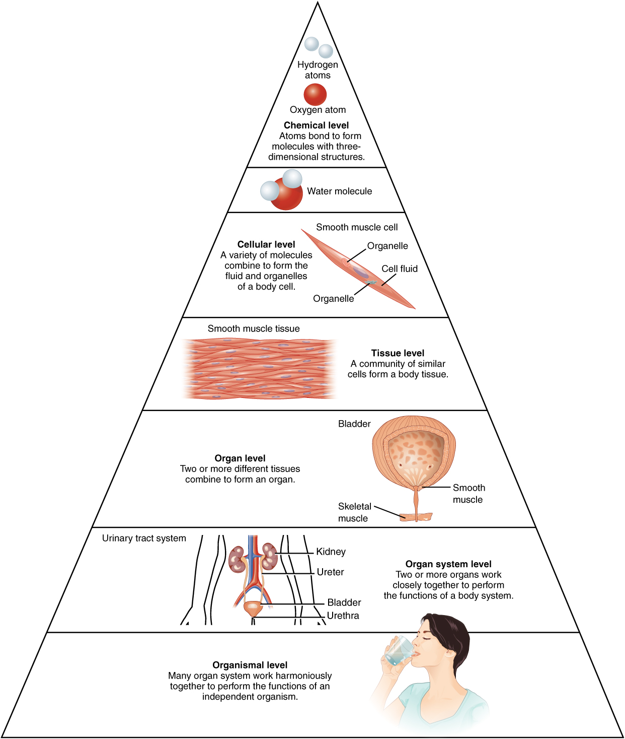
Levels of Organization
Living things are called organisms. Organisms are an organized collection of matter. The building blocks of matter are atoms. Atoms are the simplest structures that can combine to make larger things. Atoms combine to make molecules that are organized into cells. Cells are the smallest living structural units. Cells can organize into tissues. A tissue is a collection of similar cells. There are four major tissue types. When different tissues organize into a single structure it is called an organ. For instance, the heart is an organ made up of all 4 tissues: muscle, nervous, epithelial, and connective. Organs combine into organ systems. For instance, the cardiovascular system includes multiple organs such as the heart and blood vessels.

Fig. 1.6: Hierarchy of Structure
Organ Systems
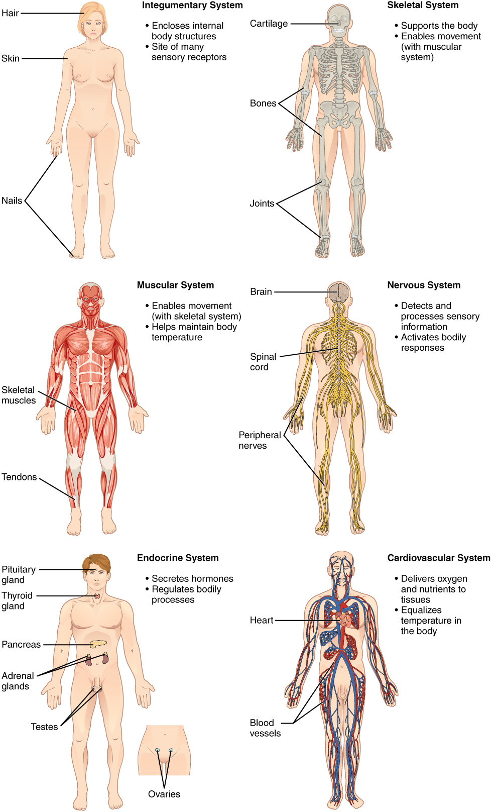
This book covers the 11 organ systems of the human body. None of these systems work completely independently of the others.
The integumentary system (skin) gives the body integrity by holding everything together. The skin is the largest organ in the body. It provides a waterproof barrier that protects against infection. The skin is also sensory (it provides information about the external environment such as touch, vibration, pain, temperature).
The skeletal system provides a framework for our bodies that enables posture and movement. It is also the site of hematopoiesis (making of blood cells) and mineral and fat storage. Osseous (bone) tissue and the skeletal system (bone organs and joints) are covered over four separate chapters.
The muscular system allows for the movement of the body and body parts. Skeletal muscles attach to bones which enable the body to move. Smooth muscles enable organs to move substances around the body such as food through the digestive tract. Cardiac (heart) muscle is unique to the heart and pumps blood throughout the body. There are two chapters covering muscles, the first is muscle tissue, and the second covers the names and actions of the major skeletal muscle organs.
The nervous system is responsible for internal and external sensation, the contraction of muscle, and excretion from endocrine and exocrine glands. Due to the complexity of this system, it is covered in five separate chapters: nervous tissue, spinal cord, brain, special senses, and the autonomic nervous system (ANS).
The endocrine system is a body-regulatory system whereby chemicals called hormones diffuse through the bloodstream and bind receptors on tissues or organs causing a physiological change.
The cardiovascular system is covered in three separate chapters: hematology (study of blood), the heart, and blood vessels. These organ systems work together to distribute nutrients (oxygen, glucose, amino acids, etc.) and remove wastes (carbon dioxide, acids, etc.) from body tissues.
The respiratory system works in conjunction with the cardiovascular system to bring oxygen into the body while removing carbon dioxide.
The immune system works to remove foreign or damaged molecules and cells from the body. This involves steps to destroy and control the spread of microorganisms and your own cells that are damaged or potentially harmful such as cancerous cells.
The digestive system is covered in two chapters. The first describes how the body works to break down foods we eat into small molecules that can be absorbed. These small molecules are used to generate energy or as building blocks to make human macromolecules. The second chapter covers cellular metabolism including the specific steps of how molecules from our food are used to generate the energy molecule ATP.
The urinary system is covered in two chapters. It removes wastes (acids, creatinine, urea) and other excess substances (water, salts, etc) from the body by filtering the blood. The first chapter covers basic anatomy and physiology of this system whereas fluid and electrolyte balance is covered in a separate chapter.
The Reproductive system enables humans to reproduce thereby propagating the species. One chapter will cover male and female reproductive anatomy and physiology. Then the book concludes with a chapter on human development which describes the processes involved in the growth and differentiation of one single cell into a mature adult.

Fig. 1.7: Organ Systems of the Human Body

Fig. 1.8: Organ Systems of the Human Body (continued)
1.3 Anatomical terminology
![]() 1.3 Learning Outcome
1.3 Learning Outcome
- Utilize anatomical position with regional and directional terminology to refer to any place on a patient’s body
Regional Names
The body axis (center line) or the axial skeleton refers to the head (cephalic), neck (cervical), and torso (thoracic and abdominal regions). The arms and legs are referred to as the upper and lower appendages (additions) and are, therefore, part of the appendicular skeleton.
Plantar and palmar refer to the bottom of the foot and anterior face of the hand, respectively. These are the more fleshy (soft tissue) sides, whereas the dorsum of the foot and the hand refers to the opposite and more bony surfaces. In other words, the dorsal and palmar surfaces of the hand are opposite one another. The dorsum of the foot is opposite of the plantar region.
The regional terms found below will be utilized in various forms, sometimes as suffixes or prefixes, in bone and muscle names, as well as other A&P and medical terms. You must learn these well as they will serve as a foundation for all future healthcare-related courses you might take. Keep in mind that this is not a complete list and any noun can be rephrased as an adjective.

Fig. 1.9: Regions of the Human Body
- Cephalic
- Cranial (brain)
- Occipital (back of the cranium, above where the head meets the neck)
- Otic/auricular (ear)
- Buccal (cheek)
- Mental (chin)
- Nasal (nose)
- Ocular/optic/orbital (eyes)
- Frontal (forehead)
- Oral (mouth)
- Maxillary (upper jaw)
- Thoracic
- Axillary (arm pit)
- Mammary (breast)
- Pectoral (chest)
- Sternal (sternum)
- Abdominopelvic
- Abdominal (stomach, belly, tummy)
- Pelvic (between the hip bones)
- Coxal (hip)
- Inguinal (groin, lower pelvic flanking the pubic)
- Pubic (genital)
- Vertebral (spine)
- Cervical (neck)
- Thoracic (rib cage)
- Lumbar (lower back)
- Sacral (where the spine meets the hips)
- Coccygeal (tailbone)
- Upper extremity
- Shoulder
- Acromial
- Glenoid
- Deltoid
- Brachial (upper arm)
- Antecubital (front of elbow)
- Olecranal (back of elbow)
- Antebrachial (forearm)
- Manus (hand)
- Dorsum (back of the hand)
- Carpal (wrist)
- Palmar (palm, front of the hand body)
- Digital (fingers)
- Shoulder
- Lower extremity
- Femoral (upper leg)
- Gluteal (buttocks)
- Patellar (knee cap)
- Popliteal (behind the knee)
- Crural (the whole lower leg)
- Sural (back of the lower leg)
- Pes (foot)
- Calcaneal (heel)
- Tarsal (ankle)
- Dorsum (top of the foot)
- Digital (toes)
- Plantar surface (sole, bottom of the foot)
![]() Retrieval Practice
Retrieval Practice
Draw a simple diagram of the human body, and label the parts of the body using the regional names above. Repeat this process until you get them all correct. Make sure to pat yourself on the back for doing this task!
Sectional Terminology

Fig. 1.10: Planes of the Body
A section is a two-dimensional surface of a three-dimensional structure that has been cut. Modern medical imaging devices enable clinicians to obtain “virtual sections” of living bodies. We call these scans. Body sections and scans can be correctly interpreted, however, only if the viewer understands the plane along which the section was made. A plane is an imaginary two-dimensional surface that passes through the body. There are three planes commonly referred to in anatomy and medicine, as illustrated in Fig 1.10.
- The sagittal plane is the plane that divides the body or an organ vertically into right and left sides. If this vertical plane runs directly down the middle of the body, it is a midsagittal or median plane. If it divides the body into unequal right and left sides, it may be called a parasagittal plane.
- The frontal plane is the plane that divides the body or an organ into anterior (front) and a posterior (rear) portions. The frontal plane is often referred to as a coronal plane (corona is Latin for crown).
- The transverse plane is the plane that divides the body or organ horizontally into upper and lower portions. Transverse planes produce images referred to as cross-sections.
- The oblique plane (not shown) is a diagonal plane that is a combination of 2 or more of the above.

Fig 1.11. images showing/using sectional terms for medical imaging such as X-rays, CT, etc
Anatomical Position and Directional Terms
The body is said to be in the prone position when it is in the anatomical position lying face down. When lying face up, it is said to be in the supine position.

Fig 1.12: Prone/supine
The anatomical position is a reference that all health professionals use with anatomical directional terminology regardless if your patient is prone, supine or in some other orientation. Notice from Fig. 1.13 that the chin and top of the foot are parallel to each other, and the arms are out to the side with the palms facing forward and thumbs pointing laterally (to the sides). When referencing anything on the body, imagine the patient inanatomical position and refer to the patient’s right and left, not yours. This ensures that in clinical situations, where the patient may be in any orientation, there won’t be any confusion when referring to the right hand, for instance.

Fig 1.13: Anatomical position
Anterior (ventral) refers to the front or towards the front. Posterior or (dorsal) refers to the back or toward the back. For example, your nose is anterior to your ears. Alternatively, your ears are dorsal to your nose.
Proximal means closer to an attachment point, whereas distal means further away. These directional terms are used only when referring to areas of the arms and legs in reference to where they attach to your body. For example, your fingers (digits) are distal to your forearm (antebrachium). The opposite is also true: your antebrachium is proximal to your digits. These terms must be used comparatively. In other words, one would never say “the elbow is proximal”. It needs to be compared to another part or region of the same limb. For example, the elbow (olecranon) is proximal to the wrist (carpal) region or the carpal region is distal to the olecranon.
Superior (cranial)/inferior (caudal) are directional terms that refer simply to up/down in the anatomical position. For example, moving closer to the head of an individual would be moving in a superior direction, whereas moving closer to the bottom of the feet would be moving in an inferior direction.
Deep/superficial refers to how far or close something is from the surface. Keep in mind the most superficial portion of the body is the top layer of the skin (or, more precisely, the hair on top of that). Deep refers to further away from the surface of the skin.
Medial/lateral refers to the midline or sides of the body. Picture drawing an imaginary line down the middle of a person facing you. This dividing line is the most medial point. If you cut them along this vertical axis, it would be considered a mid-sagittal section. As one moves towards the right or left sides the position would be lateral compared to the midline. Therefore medial is referred to as toward the midline and the lateral is away from the midline.
1.4 Body Cavities, Organs, and Membranes
![]() 1.4 Learning Outcomes
1.4 Learning Outcomes
- Identify the major body cavities and the organs they contain
- Describe the major serous membranes
- Describe the abdominal regions
Ventral

Fig. 1.14: Body Cavities
The body cavities are the spaces or compartments that house many of the body’s organs. The thoracic and abdominopelvic cavities together are known as the anterior or ventral cavity. They are separated by a distinctive border, the diaphragm muscle. The dorsal cavity (aka the posterior cavity) consists of the cranial and vertebral cavities. Both of these cavities are encased in bone to protect the delicate neural tissue of the central nervous system (CNS). The cranial cavity contains the brain whereas the vertebral cavity contains the spinal cord.

Fig 1.15: Mediastinum (heart in its space between lungs)
The thoracic cavity is on the superior side of the diaphragm muscle and consists of sub-cavities and spaces. It is divided into the right and left pleura (cavities containing the lungs) and the space in-between known as the mediastinum. The mediastinum contains major vessels that are branching out of or entering the heart as well as the trachea and esophagus. Sitting within this area is the pericardial cavity which houses the heart itself and the surrounding pericardial sac containing pericardial fluid. This fluid surrounds the heart and serves as a layer of lubrication between the heart and surrounding structures.
The abdominopelvic cavity is inferior to the thoracic cavity and diaphragm muscle. The abdominopelvic cavity can be divided into the abdominal cavity and the pelvic cavity, however a clear distinctive border does not exist here. The pelvic cavity is technically the area where the pelvic bones encircle reproductive organs and the inferior portions of the digestive tract.

Fig 1.16: Serous Membranes
The ventral body cavities and the organs within are lined by a membrane (flat, sheet-like structure) known as a serous membrane. These membranes stretch and distort to compensate for organ shape changes (i.e. a full stomach vs. empty stomach, expanding and contracting lungs). Anatomists have developed a two-part naming system for these serous membranes:
- Does the membrane surround the organ (visceral) or line the body cavity (parietal)?
- Which specific body cavity does the organ reside?
For example, in the pleural cavities, the serous membrane that surrounds the lungs is known as the visceral pleura, whereas the serous membrane that lines the pleural cavity is referred to as the parietal pleura. This naming method applies to the other ventral cavities as well. The serous membrane that surrounds the heart is called the visceral pericardium and the serous membrane that lines the pericardial cavity is called the parietal pericardium. The organs of the abdominopelvic cavity are enclosed in a serous membrane called the peritoneum. It blankets the abdominopelvic organs (visceral peritoneum) and forms the inner face of the outer abdominopelvic wall (parietal peritoneum).
Cells of serous membranes secrete a liquid known as serous fluid into the space between the visceral and parietal layers. This fluid lubricates and protects organs by reducing friction and heat caused by movement of internal body parts such as heart, lungs and digestive tract.
Let’s compare serous membranes to an avocado peel. Pretend an avocado is an organ in the abdomen. If you remove the peel and touch the outer part of the avocado flesh you are touching the visceral (viscera = internal organs) peritoneum. If you then touch the inside of the peel you are now touching the parietal peritoneum. If the space between the two membranes was filled with a fluid you could imagine that maybe your avocados wouldn’t bruise so easily. That’s the protective function of serous fluid.
Now imagine the outer avocado flesh folded outwards to create the peel. The visceral and parietal serous membrane layers are indeed folds of the same tissue. The visceral pericardium folds outwards creating the parietal pericardium. Similarly, the visceral and parietal peritoneum are also folds of the same tissue. The peritoneum also folds and thickens in places to anchor and nourish the organs of the abdominal cavity. This idea of a membrane folding to create other membranes and more elaborate structures is a common theme in anatomy and will be revisited in other chapters.
The abdominopelvic cavity has a collection of organs that sit behind the peritoneal cavity which is known as the retroperitoneal space. Organs in this space either have only one side covered in peritoneum or none at all. Organs that reside within this space include the kidneys, portions of the upper GI tract, and the pancreas.

Fig 1.17: [Retroperitoneum (retroperitoneal image)]

Fig 1.18: Regions
In addition to directional terminology, clinicians and anatomists will also use abdominal regional terms. This allows them to refer to more precise areas of this cavity that contains many organs. Abdominopelvic regions map out the abdomen and pelvic area into nine subsections (Figure 1.18, A). Keep in mind these aren’t separated from each other anatomically, you can see in the picture that individual organs occupy space in more than one region.
Another way of dividing the abdomen is into 4 areas known as abdominopelvic quadrants. These quadrants are made by crossing horizontal and vertical lines that intersect at the umbilicus (belly button or piko in Hawaiian). Therefore the two upper quadrants are known as the right and left upper quadrants and the two lower quadrants are known as the right and left lower quadrants. This system is more commonly used by nurses and physicians.
1.5 Homeostasis
![]() 1.5 Learning Outcomes
1.5 Learning Outcomes
- Define homeostasis
- Contrast negative and positive feedback systems
- Explain the scientific method
Homeo- (similar) stasis (inactive, not moving or changing) is the process of maintaining a constant physiological state. In reality, our bodies are always changing and homeostatic processes are the mechanisms used to readjust to maintain a healthy physiology. The anatomical structures involved in maintaining homeostasis are sensors (sensory receptors), a control center (often an area of the brain), and effectors (a target organ or tissue that changes in response to the stimulus). Sensors that change in response to stimuli send a signal to the control center. Then the control center sends a signal to effector organs such as skeletal muscles.

Fig 1.19: Illustration of the receptor, control center, and effector
To better understand homeostasis, we can use ahupuaʻa land management system as a metaphor. This traditional Hawaiʻian land management system is a section of land spanning mauka (mountain) to makai (sea). Each ahupuaʻa encompassed different geologic and climate zones that provided the necessary foods and materials for people within that area to be self-sustainable. Different groups of people cared for different sections, and they traded what they couldnʻt make or get for themselves with others. To maintain balance, resources that were limited were conserved by placing them under kapu (forbidden, prohibited). In traditional fishing practice, when one beach or fishing ground (i.e. deep sea fishing or coral fishing) was kapu, another was open. Kapu ensured that the marine ecosystem Hawaiians depended so greatly on was balanced. In much the same manner, different systems of the body respond to internal and external environmental changes by conserving or producing resources until the body can return to its normal, healthy state.

Fig 1.20: Oʻahu Ahupuaʻa
An example of homeostatic maintenance is that of body temperature. In this example, body temperature is the controlled condition that needs to be in balance. If your body gets too hot, it tries to cool down through perspiration (sweating). If your body temperature drops too low, the body will try to warm up by shivering. This is an example of negative feedback regulation and describes most homeostatic processes.

Fig. 1.21: Negative Feedback Loop
A negative feedback system reverses deviation from a set point, a physiological value or range by which your body physiology tries to maintain. Blood glucose concentration is another example of a negative feedback system. If blood glucose is too high, insulin will be released which causes glucose concentrations to drop. However, if the concentrations are too low, glucagon will be released which causes glucose concentrations to rise. In each of these cases, the body tries to maintain a set point by responding to extremes (too much or too little of something) and then negative feedback processes bring that value back within a homeostatic range.
Due to negative feedback mechanisms, healthy physiology can be maintained. The vast majority of homeostatic processes involve negative feedback. Since numerous physiological parameters of various people in various states of health have been determined, certain ranges can be associated with not only health but specific pathologies (disease). Therefore, health care professionals evaluate these parameters to determine the cause of pathology. It is generally assumed, on average, that around 95% of healthy people will fall into these normal ranges.
Physiology value ranges for various physiological parameters of healthy adults.
- Heart Rate: 60-80 beats per minute
- Glucose: 80-110 mg/dL
- Body Temperature: 98.1-98.9o F (36.7-37.2o C)
- Breathing Rate: 15-20 breaths per minute

Fig 1.22: Positive feedback loop
There are a few examples of positive feedback whereby a change in a controlled condition is strengthened or reinforced. For instance, when a blood vessel is damaged, platelets are recruited to plug the hole to stop the hemorrhaging (bleeding). This process is called hemostasis (hemo, blood; stasis, not changing). The act of one platelet binding a damaged capillary stimulates additional platelets to bind. Once the bleeding stops, other mechanisms trigger to stop the clotting process. Otherwise, blood vessels would become blocked preventing blood flow.
Oxytocin is a hormone with numerous effects around the body. During childbirth, the fetus’ head stretches the smooth muscle of the cervix which is part of the uterus. This stimulus causes oxytocin release from the mother’s brain. Oxytocin diffuses through the blood, binds receptors on smooth muscle cells causing the uterus to contract which allows the baby’s head to further stretch the cervix. This increases additional oxytocin release which in turn intensifies uterine wall contractions. This positive feedback continues until the baby is delivered and the cervix is no longer being stretched.
Another role of the oxytocin hormone is the milk let-down reflex. A baby suckling on the breast stimulates mammary sensory receptors to send a signal to the brain causing the release of oxytocin. Oxytocin diffuses through the blood, eventually binding mammary receptors resulting in the release of milk. As long as suckling continues regularly, milk will continue to be released.
Homeostatic imbalances occur when physiological processes fail to maintain parameters within healthy ranges. For instance, diabetes is a condition whereby an individual cannot maintain blood glucose within a healthy range. This may lead to other health issues such as stroke or kidney failure. If normal physiology cannot be maintained, depending on the pathology, possible treatments include removing the source of injury, lifestyle change, therapy (a routine exercise), drugs, or surgery. Generally, drugs are used to either mimic or replace a molecule of your body or block one molecule from binding another.
![]() Cultural Connection
Cultural Connection
Lōkahi and homeostasis. Lo- (to obtain), -kahi (one unit). Lōkahi is a Hawaiian value that can be associated with unity, and hoʻo lōkahi is a reference to the ability to get or obtain balance or be in agreement. In Hawaiian culture, the Lōkahi wheel represents six sections of the life of a kanaka (person): Spiritual/Soul, Friends/Family, Physical/Body, Work/School, Thinking/Mind, and Feelings/Emotions. Each of the six sections must be balanced to make one big wheel that represents the person. We also need to know what each section looks like for us, to understand how to establish balance in our lives.
 Fig 1.23 Figure of Lōkahi wheel
To determine the cause of disease, scientists use the scientific method. This is a series of steps that should be followed by all scientists to maintain the integrity of scientific discovery. The scientific method steps are:
- Make observations
- Form hypotheses and null hypotheses (testable statements)
- Design and execute experiments
- Analyze data
- Compare results to the null hypothesis
Making observations may include looking at patients’ physiological values, asking questions regarding health and lifestyle history or considering the results of previous research. Based on these observations, we can form testable questions. For instance, after observing many patients you may think there is a link between diabetes and kidney failure. Then you form your hypothesis: diabetes causes kidney failure. This statement is called the hypothesis. The null hypothesis is the opposite statement: diabetes doesn’t cause kidney failure. Depending on the hypothesis, designing good experiments isn’t always easy and may require creativity and cleverness on the part of the scientist. Data analysis often involves quantitative reasoning including statistical analysis. Finally, the results of the analysis are compared to the null hypothesis (not the hypothesis). This prevents individual scientists from claiming they have proven anything in particular. In other words, you can’t prove your hypothesis. At best you can only fail to deny the null hypothesis. This is meant to ensure the scientific field as a whole continues to move closer to the truth by adhering to the premise that no one individual or group can ever attain it.

Chapter Summary
Quiz

Sources
Nana i ke kumu (Look to the source). Vol.1 Mary Kawena Pukui, EW Haertig, M.D., Catherine A. Lee, Queen Liliʻuokalani Childrenʻs Center from Ulukau: The Hawaiian Electronic Library
Nā Puke Ulukau Website
Polynesian Voyaging Society
Founding the Polynesian Voyaging Society; Building and Naming Hōkūle‘a
In Search of the Ancient Polynesian Voyaging Canoe (1998)
The Building of the Hokule‘a – 1973-75
Ahupuaʻa
Kumakahi: Living Hawaiian Culture Website
Lokahi
E Malāma Pono Website
Ho’okua’āina Website
Key Terms
abdominal
relating to the abdomen, the superior portion of the abdominopelvic cavity
abdominal cavity
the space bounded by the abdominal walls, diaphragm, and pelvis
abdominopelvic cavity
division of the anterior (ventral) cavity that houses the abdominal and pelvic viscera
anatomical position
standard reference position used for describing locations and directions on the human body
anatomy
science that studies the form and composition of the body’s structures
anterior
describes the front or direction toward the front of the body; also referred to as ventral
anterior cavity
larger body cavity located anterior to the posterior (dorsal) body cavity; includes the serous membrane-lined pleural cavities for the lungs, pericardial cavity for the heart, and peritoneal cavity for the abdominal and pelvic organs; also referred to as ventral cavity
atoms
the simplest form of matter that can be combined to make molecules
caudal
describes a position below or lower than another part of the body proper; near or toward the tail (in humans, the coccyx, or lowest part of the spinal column); also referred to as inferior
cell
the basic structural unit of all organisms
cephalic
relating to the head
cervical
relating to the neck
control center
compares values to their normal range; deviations cause the activation of an effector
cranial
describes a position above or higher than another part of the body proper; also referred to as superior
cranial cavity
division of the posterior (dorsal) cavity that houses the brain
deep
describes a position farther from the surface of the body
diaphragm
the partition separating the thoracic cavity from the abdominal cavity
differentiation
process by which unspecialized cells become specialized in structure and function
distal
describes a position farther from the point of attachment or the trunk of the body
dorsal
describes the back or direction toward the back of the body; also referred to as posterior
dorsal cavity
posterior body cavity that houses the brain and spinal cord; also referred to the posterior body cavity
effector
organ that can cause a change in a value
etymology
the derivation of a word or word history
frontal plane
two-dimensional, vertical plane that divides the body or organ into anterior and posterior portions
gross anatomy
study of the larger structures of the body, typically with the unaided eye; also referred to as macroscopic anatomy
growth
process of increasing in size
homeostasis
steady state of body systems that living organisms maintain
inferior
describes a position below or lower than another part of the body proper; near or toward the tail (in humans, the coccyx, or lowest part of the spinal column); also referred to as caudal
lateral
describes the side or direction toward the side of the body
macroscopic anatomy
study of the larger structures of the body, typically with the unaided eye; also referred to as gross anatomy
medial
describes the middle or direction toward the middle of the body
metabolism
sum of all of the body’s chemical reactions
microscopic anatomy
study of very small structures of the body using magnification
molecule
an organized structure of bonded atoms
movement
the change of position by an organism or part of organism, often in response to stimuli
negative feedback
homeostatic mechanism that tends to stabilize an upset in the body’s physiological condition by preventing an excessive response to a stimulus, typically as the stimulus is removed
normal range
range of values around the set point that do not cause a reaction by the control center
oblique plane
neither perpendicular nor parallel to a given line or surface; slanting; sloping
organ
functionally distinct structure composed of two or more types of tissues
organ system
group of organs that work together to carry out a particular function
organism
living being that has a cellular structure and that can independently perform all physiologic functions necessary for life
organization
the specific arrangement of atoms, molecules, cells, tissues and organs that define an organism
palmar
of, relating to, or located in or on the palm of the hand
parietal pericardium
the serous membrane lining the pericardial cavity
parietal peritoneum
the serous membrane lining the abdominal cavity
parietal pleura
the serous membrane lining the pleural cavities
pelvic cavity
the space bounded by the bones of the pelvis and pelvic girdle
pericardial cavity
the cavity encasing the heart
pericardium
sac that encloses the heart
peritoneum
serous membrane that lines the abdominopelvic cavity and covers the organs found there
physiology
science that studies the chemistry, biochemistry, and physics of the body’s functions
plantar
of or relating to the sole of the foot
plane
imaginary two-dimensional surface that passes through the body
pleura
serous membrane that lines the pleural cavity and covers the lungs
positive feedback
mechanism that intensifies a change in the body’s physiological condition in response to a stimulus
posterior
describes the back or direction toward the back of the body; also referred to as dorsal
posterior cavity
posterior body cavity that houses the brain and spinal cord; also referred to as dorsal cavity
prone
face down
proximal
describes a position nearer to the point of attachment or the trunk of the body
regulation
the process of maintaining a physiological equilibrium, see homeostasis
repetition
the practice of going through learning materials several times as part of the studying process
reproduction
process by which new organisms are generated
responsiveness
ability of an organisms or a system to adjust to changes in conditions
sagittal plane
two-dimensional, vertical plane that divides the body or organ into right and left sides
scientific method
the process of scientific discovery whereby observations lead to the formation of hypotheses, experiments are designed and performed, and data are compared to null hypotheses for drawing conclusions
section
in anatomy, a single flat surface of a three-dimensional structure that has been cut through
self-quizzing
the practice of testing oneself as part of the studying process
sensor
(also, receptor) reports a monitored physiological value to the control center
serous fluid
A lubricating fluid produced by and between folds of serous membrane
serous membrane
membrane that covers organs and reduces friction; also referred to as serosa
set point
ideal value for a physiological parameter; the level or small range within which a physiological parameter such as blood pressure is stable and optimally healthful, that is, within its parameters of homeostasis
superficial
describes a position nearer to the surface of the body
superior
describes a position above or higher than another part of the body proper; also referred to as cranial
supine
face up
thoracic
relating to the superior ventral cavity; the thoracic cavity
thoracic cavity
division of the anterior (ventral) cavity that houses the heart, lungs, esophagus, and trachea
tissue
an aggregate of similar cells and cell products forming a definite kind of structural material
transformation
to change from one form to another; as a study strategy this refers to the practice of transforming the information in narrative form to a diagram, flow chart or other illustration
transverse plane
two-dimensional, horizontal plane that divides the body or organ into superior and inferior portions
ventral
describes the front or direction toward the front of the body; also referred to as anterior
ventral cavity
larger body cavity located anterior to the posterior (dorsal) body cavity; includes the serous membrane-lined pleural cavities for the lungs, pericardial cavity for the heart, and peritoneal cavity for the abdominal and pelvic organs; also referred to as anterior body cavity
visceral pericardium
the serous membrane covering the heart
visceral peritoneum
the serous membrane covering the abdominal cavity viscera
visceral pleura
the serous membrane covering the lungs
X-ray
form of high energy electromagnetic radiation with a short wavelength capable of penetrating solids and ionizing gases; used in medicine as a diagnostic aid to visualize body structures such as bones
Media Attributions
- olelo_noeau
- Hokule’a.jpg © Phil Uhl is licensed under a CC BY (Attribution) license
- Anatomy & Physiology © LynleyShimat Lys, OpenStax, Yathin sk is licensed under a CC BY-SA (Attribution ShareAlike) license
- HawaiianBoatDiagram © Tyler English is licensed under a CC BY (Attribution) license
- Gross and microscopic anatomy © Openstax, Regents of University of Michigan Medical School is licensed under a CC BY (Attribution) license
- woman writing on book © Kyle Gregory Devaras is licensed under a CC0 (Creative Commons Zero) license
- Levels of Structural Organization of the Human Body © Openstax is licensed under a CC BY (Attribution) license
- Organ Systems Human Body © Openstax is licensed under a CC BY (Attribution) license
- Organ Systems of the Human Body (continued) © Openstax is licensed under a CC BY (Attribution) license
- Regions of the Human Body © Openstax is licensed under a CC BY (Attribution) license
- Planes of the Body © Openstax is licensed under a CC BY (Attribution) license
- Medical_Imaging_Techniques © Oregon State University is licensed under a CC BY-SA (Attribution ShareAlike) license
- Supine position and prone position © Jmarchn is licensed under a CC BY-SA (Attribution ShareAlike) license
- Directional Terms Applied to the Human Body © Openstax is licensed under a CC BY (Attribution) license
- Dorsal and Ventral
- Annotated version of Gray’s image 969; opened Mediastinum viewed from left body side © Wikimedia Commons is licensed under a Public Domain license
- Serous Membranes © Openstax is licensed under a CC BY (Attribution) license
- Retroperitoneal_spaces © Goran Mitreski and Tom Sutherland is licensed under a CC BY (Attribution) license
- Regions and Quadrants of the Peritoneal Cavity © Openstax is licensed under a CC BY (Attribution) license
- Negative_Feedback_Loops © Openstax is licensed under a CC BY (Attribution) license
- Closer view of the ahupuaʻa of Oʻahu’s south and east side © KarlM is licensed under a CC BY-SA (Attribution ShareAlike) license
- Negative_Feedback_Loops © Openstax is licensed under a CC BY (Attribution) license
- Positive Feedback Loop © Openstax is licensed under a CC BY (Attribution) license
- Lōkahi Wheel © LynleyShimat Lys is licensed under a CC BY (Attribution) license
- Honu_‘Iwalani Clayton_CCBY_2022 10 30 © ‘Iwalani Clayton is licensed under a CC BY (Attribution) license
- divider_maile
The specific arrangement of atoms, molecules, cells, tissues and organs that define an organism.
Functionally distinct structure composed of two or more types of tissues.
Standard reference position used for describing locations and directions on the human body.
science that studies the form and composition of the body’s structures
science that studies the chemistry, biochemistry, and physics of the body’s functions
form of high energy electromagnetic radiation with a short wavelength capable of penetrating solids and ionizing gases; used in medicine as a diagnostic aid to visualize body structures such as bones
The basic structural unit of all organisms.
An aggregate of similar cells and cell products forming a definite kind of structural material.
Two or more atoms covalently bonded together.
Study of the larger structures of the body, typically with the unaided eye; also referred to as macroscopic anatomy.
Study of the larger structures of the body, typically with the unaided eye; also referred to as gross anatomy.
Study of very small structures of the body using magnification.
Group of organs that work together to carry out a particular function.
The derivation of a word or word history.
In anatomy, a single flat surface of a three-dimensional structure that has been cut through.
The practice of going through learning materials several times as part of the studying process.
To change from one form to another; as a study strategy this refers to the practice of transforming the information in narrative form to a diagram, flow chart or other illustration.
The practice of testing oneself as part of the studying process.
Sum of all of the body’s chemical reactions.
Process of increasing in size.
Process by which unspecialized cells become specialized in structure and function.
Ability of an organisms or a system to adjust to changes in conditions.
The change of position by an organism or part of organism, often in response to stimuli.
Living being that has a cellular structure and that can independently perform all physiologic functions necessary for life.
The process of maintaining a physiological equilibrium, see homeostasis.
Process by which new organisms are generated.
The simplest form of matter that can be combined to make molecules.
Relating to the head.
Relating to the neck.
Relating to the superior ventral cavity; the thoracic cavity.
Relating to the abdomen, the superior portion of the abdominopelvic cavity.
Of or relating to the sole of the foot.
Of, relating to, or located in or on the palm of the hand.
Describes the front or direction toward the front of the body; also referred to as ventral.
Describes the back or direction toward the back of the body; also referred to as posterior.
Describes a position above or higher than another part of the body proper; also referred to as superior.
Imaginary two-dimensional surface that passes through the body.
Two-dimensional, vertical plane that divides the body or organ into right and left sides.
Two-dimensional, vertical plane that divides the body or organ into anterior and posterior portions.
Describes the back or direction toward the back of the body; also referred to as dorsal.
Two-dimensional, horizontal plane that divides the body or organ into superior and inferior portions.
Neither perpendicular nor parallel to a given line or surface; slanting; sloping.
Face down.
Face up.
Describes the front or direction toward the front of the body; also referred to as anterior.
Describes a position nearer to the point of attachment or the trunk of the body.
Describes a position farther from the point of attachment or the trunk of the body.
Describes a position above or higher than another part of the body proper; also referred to as cranial.
Describes a position below or lower than another part of the body proper; near or toward the tail (in humans, the coccyx, or lowest part of the spinal column); also referred to as inferior.
Describes a position below or lower than another part of the body proper; near or toward the tail (in humans, the coccyx, or lowest part of the spinal column); also referred to as caudal.
Describes a position farther from the surface of the body.
Describes a position nearer to the surface of the body.
Describes the middle or direction toward the middle of the body.
Describes the side or direction toward the side of the body.
Membrane that covers organs and reduces friction; also referred to as serosa.
Larger body cavity located anterior to the posterior (dorsal) body cavity; includes the serous membrane-lined pleural cavities for the lungs, pericardial cavity for the heart, and peritoneal cavity for the abdominal and pelvic organs; also referred to as anterior body cavity.
the partition separating the thoracic cavity from the abdominal cavity
Posterior body cavity that houses the brain and spinal cord; also referred to the posterior body cavity.
Posterior body cavity that houses the brain and spinal cord; also referred to as dorsal cavity.
Interior space of the skull that houses the brain.
Division of the anterior (ventral) cavity that houses the heart, lungs, esophagus, and trachea.
The cavity encasing the heart.
Division of the anterior (ventral) cavity that houses the abdominal and pelvic viscera.
The space bounded by the abdominal walls, diaphragm, and pelvis.
The space bounded by the bones of the pelvis and pelvic girdle.
Serous membrane that lines the pleural cavity and covers the lungs.
The serous membrane covering the lungs.
The serous membrane lining the pleural cavities.
Sac that encloses the heart.
The serous membrane lining the pericardial cavity.
Serous membrane that lines the abdominopelvic cavity and covers the organs found there.
The serous membrane covering the abdominal cavity viscera.
The serous membrane lining the abdominal cavity.
A lubricating fluid produced by and between folds of serous membrane.
The serous membrane covering the heart.
Steady state of body systems that living organisms maintain.
Mechanism that intensifies a change in the body’s physiological condition in response to a stimulus.
The process of scientific discovery whereby observations lead to the formation of hypotheses, experiments are designed and performed, and data are compared to null hypotheses for drawing conclusions.
(Also, receptor) reports a monitored physiological value to the control center.
Compares values to their normal range; deviations cause the activation of an effector.
Organ that can cause a change in a value.
Homeostatic mechanism that tends to stabilize an upset in the body’s physiological condition by preventing an excessive response to a stimulus, typically as the stimulus is removed.
Ideal value for a physiological parameter; the level or small range within which a physiological parameter such as blood pressure is stable and optimally healthful, that is, within its parameters of homeostasis.
Range of values around the set point that do not cause a reaction by the control center.
